















©The Author(s) 2022.
World J Clin Cases. Aug 6, 2022; 10(22): 7950-7959
Published online Aug 6, 2022. doi: 10.12998/wjcc.v10.i22.7950
Published online Aug 6, 2022. doi: 10.12998/wjcc.v10.i22.7950
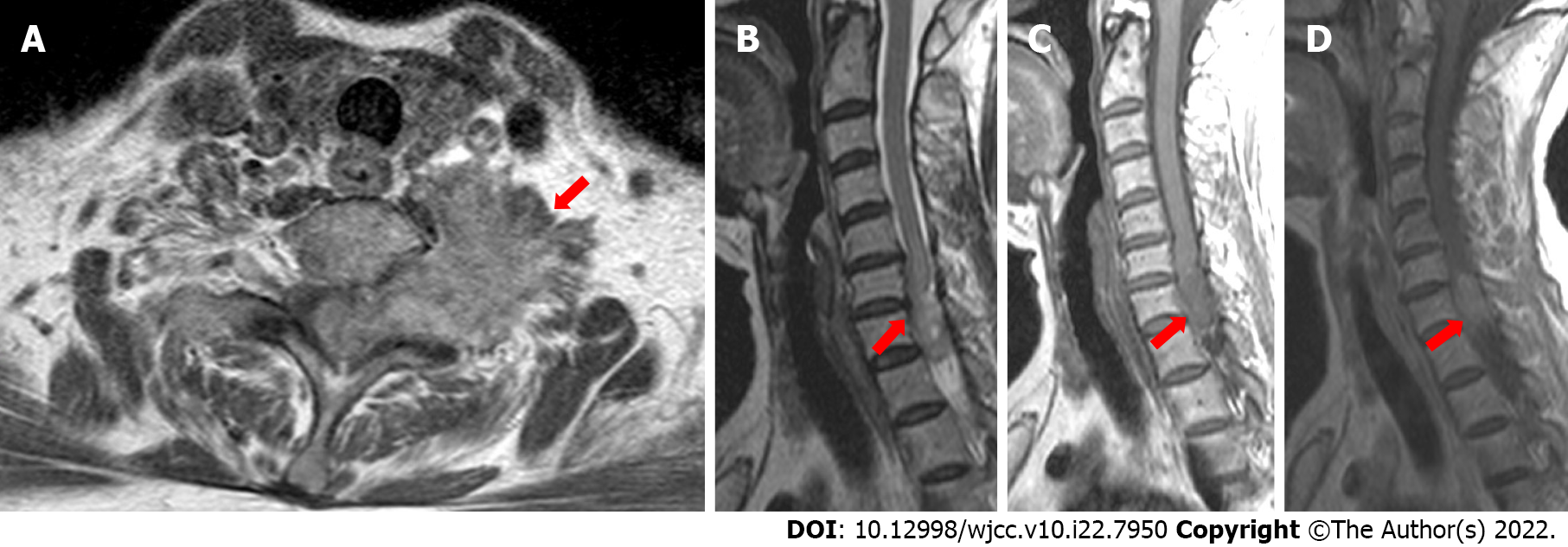
Figure 3 Magnetic resonance images of the present case.
A-C: The tumor was irregular in shape with a rough margin, and having slightly lower signal intensity on T1-weighted imaging and slightly higher signal intensity on T2-weighted imaging; D: The mass had heterogeneous enhancement.
- Citation: Liang XY, Chen YP, Li Q, Zhou ZW. Atypical imaging features of the primary spinal cord glioblastoma: A case report. World J Clin Cases 2022; 10(22): 7950-7959
- URL: https://www.wjgnet.com/2307-8960/full/v10/i22/7950.htm
- DOI: https://dx.doi.org/10.12998/wjcc.v10.i22.7950