Copyright
©The Author(s) 2022.
World J Clin Cases. Jul 26, 2022; 10(21): 7592-7598
Published online Jul 26, 2022. doi: 10.12998/wjcc.v10.i21.7592
Published online Jul 26, 2022. doi: 10.12998/wjcc.v10.i21.7592
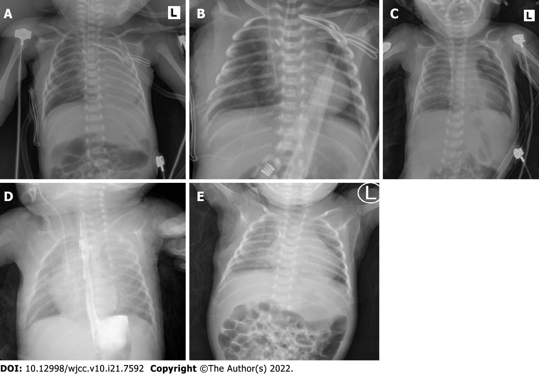
Figure 3 The chest X-ray images.
A: The chest X-ray revealed partial atelectasis of the left lung after the operation; B: The chest X-ray taken on the first postoperative day revealed good re-expansion of the left lung; C: The chest X-ray taken on the 13th postoperative day revealed pneumothorax reappearing on the left; D: The postoperative upper gastrointestinal radiography showed that the operation effect was good; E: The chest X-ray before discharge showed marked improvement.
- Citation: Zhang X, Song HC, Wang KL, Ren YY. Considerations of single-lung ventilation in neonatal thoracoscopic surgery with cardiac arrest caused by bilateral pneumothorax: A case report. World J Clin Cases 2022; 10(21): 7592-7598
- URL: https://www.wjgnet.com/2307-8960/full/v10/i21/7592.htm
- DOI: https://dx.doi.org/10.12998/wjcc.v10.i21.7592


