Copyright
©The Author(s) 2022.
World J Clin Cases. Jul 16, 2022; 10(20): 7147-7152
Published online Jul 16, 2022. doi: 10.12998/wjcc.v10.i20.7147
Published online Jul 16, 2022. doi: 10.12998/wjcc.v10.i20.7147
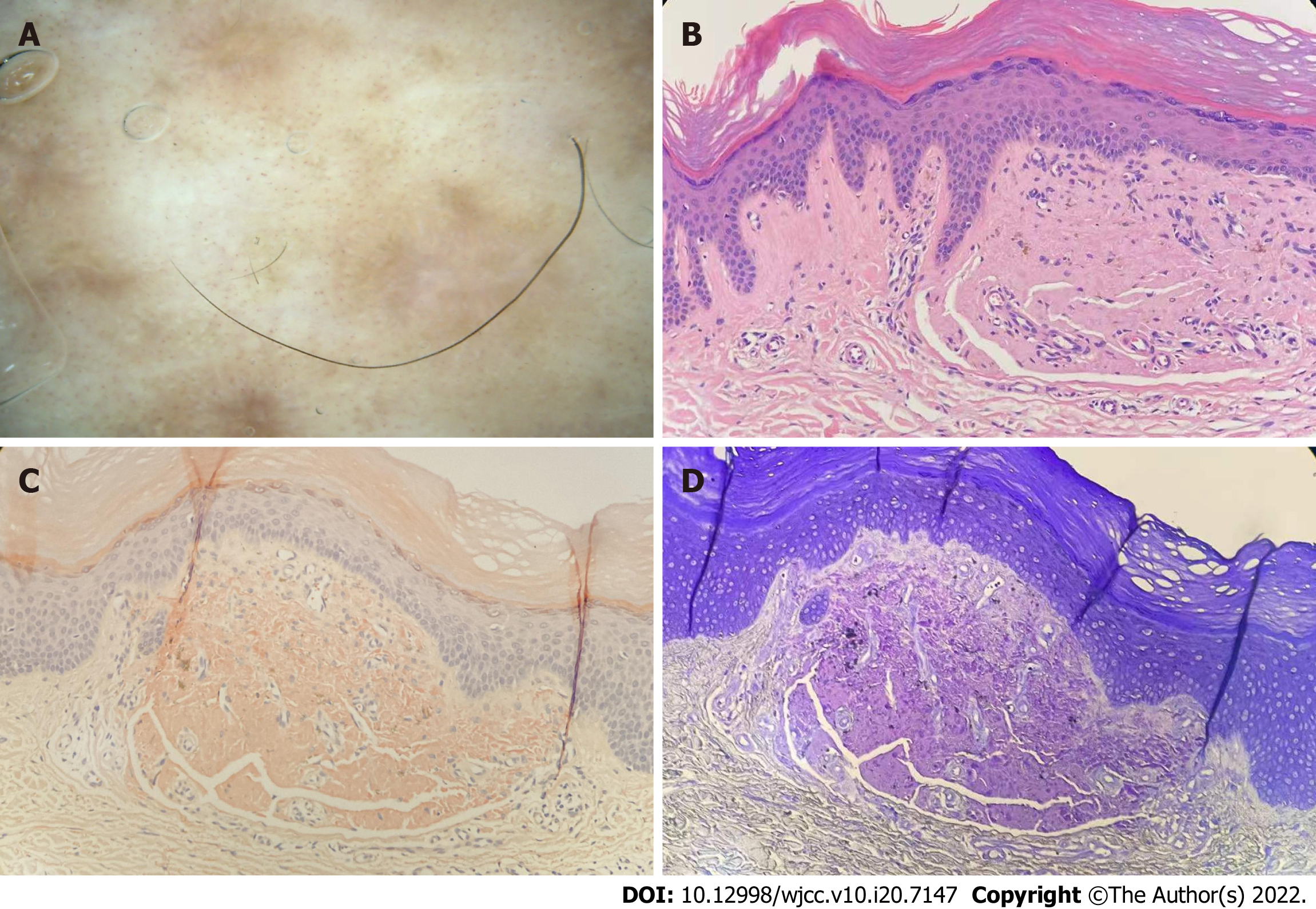
Figure 3 Dermatoscopic and dermatopathological findings in case 1.
A: The epidermis was hyperkeratinized. There were homogeneous red-stained amorphous substances in the dermis. Lymphocytes were scattered around the blood vessels; B: Amorphous substance was dyed orange; C: Amorphous substance was dyed purple; D: Dermatoscopy showed that the lesions were evenly distributed with gray pigmentation radially and dotted blood vessels densely distributed around them, and the central area was light white or light red without structure.
- Citation: Su YQ, Liu ZY, Wei G, Zhang CM. Topical halometasone cream combined with fire needle pre-treatment for treatment of primary cutaneous amyloidosis: Two case reports. World J Clin Cases 2022; 10(20): 7147-7152
- URL: https://www.wjgnet.com/2307-8960/full/v10/i20/7147.htm
- DOI: https://dx.doi.org/10.12998/wjcc.v10.i20.7147


