
















©The Author(s) 2022.
World J Clin Cases. Jan 14, 2022; 10(2): 725-732
Published online Jan 14, 2022. doi: 10.12998/wjcc.v10.i2.725
Published online Jan 14, 2022. doi: 10.12998/wjcc.v10.i2.725
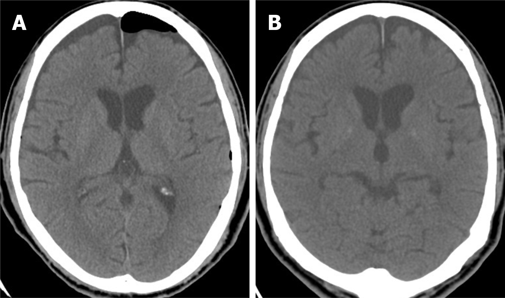
Figure 5 Post-operative computed tomography images demonstrate successful repair.
A-B: Axial view of brain computed tomography on the 5th (A) and 15th postoperative day (B) showing resolution of pneumocephalus and pneumoventricle though subdural effusion accumulation without brain parenchymal compression.
- Citation: Chang CY, Hung CC, Liu JM, Chiu CD. Tension pneumocephalus following endoscopic resection of a mediastinal thoracic spinal tumor: A case report. World J Clin Cases 2022; 10(2): 725-732
- URL: https://www.wjgnet.com/2307-8960/full/v10/i2/725.htm
- DOI: https://dx.doi.org/10.12998/wjcc.v10.i2.725