Copyright
©The Author(s) 2022.
World J Clin Cases. Jan 14, 2022; 10(2): 663-670
Published online Jan 14, 2022. doi: 10.12998/wjcc.v10.i2.663
Published online Jan 14, 2022. doi: 10.12998/wjcc.v10.i2.663
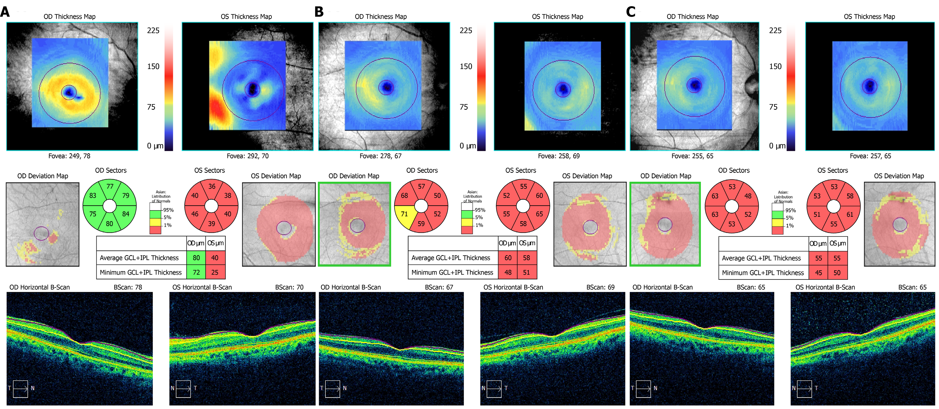
Figure 5 Ganglion cell layer and inner plexiform layer high-definition optical coherence tomography.
A: At the first visit, the thickness of ganglion cell layer and inner plexiform layer (GCIPL) in the right eye was normal, and the GCIPL in the left eye was significantly lower than normal; B: At the second visit, the GCIPL of the right eye also decreased; C: At the third visit, the binocular visual function recovered, but the GCIPL was still lower than normal without any obvious recovery.
- Citation: Sheng WY, Wu SQ, Su LY, Zhu LW. Ethambutol-induced optic neuropathy with rare bilateral asymmetry onset: A case report. World J Clin Cases 2022; 10(2): 663-670
- URL: https://www.wjgnet.com/2307-8960/full/v10/i2/663.htm
- DOI: https://dx.doi.org/10.12998/wjcc.v10.i2.663


