Copyright
©The Author(s) 2022.
World J Clin Cases. Jun 26, 2022; 10(18): 6148-6155
Published online Jun 26, 2022. doi: 10.12998/wjcc.v10.i18.6148
Published online Jun 26, 2022. doi: 10.12998/wjcc.v10.i18.6148
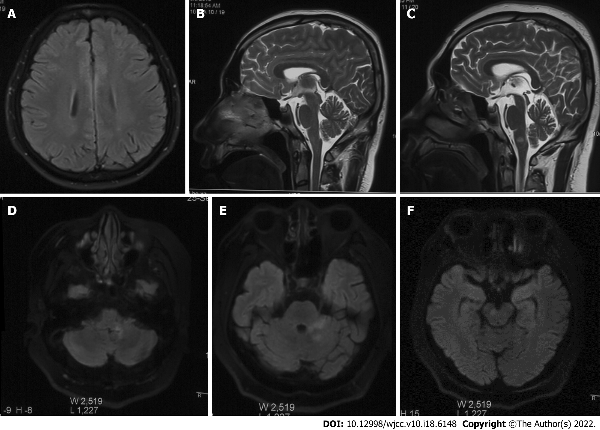
Figure 1 Imaging changes in the pathogenesis of overlapping syndrome.
A: Punctate abnormality in left parietal lobe (first episode); B: Normal sagittal position; C: High signal intensity was identified in the medulla oblongata in the T2 sagittal view; D: High signal intensity was identified in the left medulla oblongata and cerebellum of Flair; E: Flair showed hyperintensity in the left pontine arm and left cerebellum; F: Flair showed hyperintensity in the right midbrain.
- Citation: Yin XJ, Zhang LF, Bao LH, Feng ZC, Chen JH, Li BX, Zhang J. Overlapping syndrome of recurrent anti-N-methyl-D-aspartate receptor encephalitis and anti-myelin oligodendrocyte glycoprotein demyelinating diseases: A case report. World J Clin Cases 2022; 10(18): 6148-6155
- URL: https://www.wjgnet.com/2307-8960/full/v10/i18/6148.htm
- DOI: https://dx.doi.org/10.12998/wjcc.v10.i18.6148


