Copyright
©The Author(s) 2022.
World J Clin Cases. Jun 16, 2022; 10(17): 5884-5892
Published online Jun 16, 2022. doi: 10.12998/wjcc.v10.i17.5884
Published online Jun 16, 2022. doi: 10.12998/wjcc.v10.i17.5884
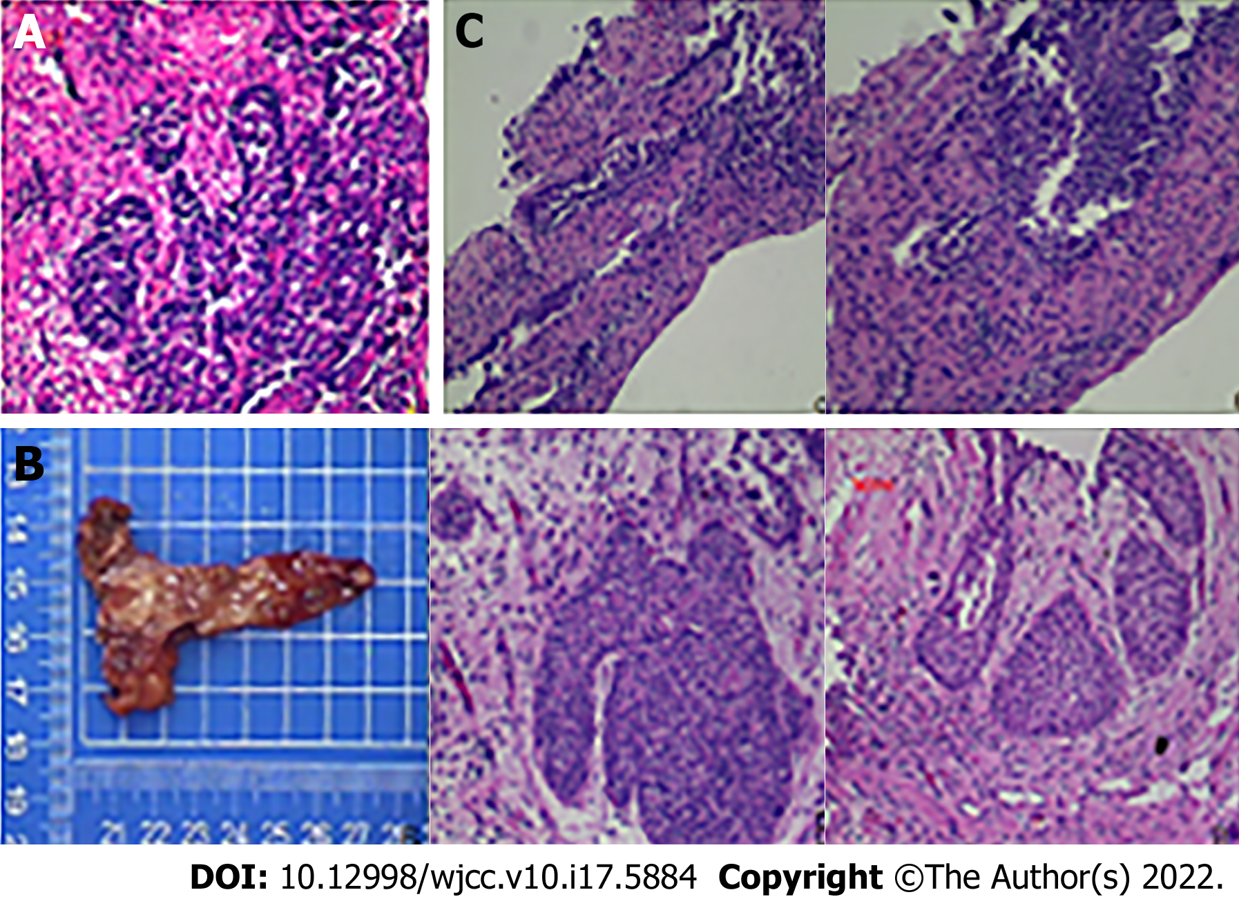
Figure 1 Postoperative pathology in 2016 and 2019.
A: After ureteral biopsy in 2016. Carcinoma tissue was suspected. Under the microscope, mucosal tissue was mixed with hemorrhagic necrotic tissue, and the surface of the mucosa was covered with urothelium, which showed papillary or solid nest like inverted growth. The tumor nucleus was oval, the cytoplasm was deep, mitosis was occasionally seen, and the focal area seemed to be a staggered arrangement of solid nestlike cells and proliferative stroma; B: After ureteral bladder replantation. Chronic mucositis and mild atypical hyperplasia was considered. Under the microscope, the urinary tract epithelium covered by the mucosal surface of some areas proliferated, and grew to the lamina propria to form a small nest or glandular tube-like structure, focal squamous metaplasia, partial mucosal surface necrosis, covered with a large amount of red staining without structure, and significant interstitial edema in the lamina propria; C: after ureteral biopsy in 2019. Polypoid change was showed. Under the microscope, part of the surface is lined with hyperplastic urothelial epithelium and lamina propria fibrous interstitial hyperplasia.
- Citation: Xie K, Li XY, Liao BJ, Wu SC, Chen WM. Primary renal small cell carcinoma: A case report. World J Clin Cases 2022; 10(17): 5884-5892
- URL: https://www.wjgnet.com/2307-8960/full/v10/i17/5884.htm
- DOI: https://dx.doi.org/10.12998/wjcc.v10.i17.5884


