
















©The Author(s) 2022.
World J Clin Cases. Jun 16, 2022; 10(17): 5776-5782
Published online Jun 16, 2022. doi: 10.12998/wjcc.v10.i17.5776
Published online Jun 16, 2022. doi: 10.12998/wjcc.v10.i17.5776
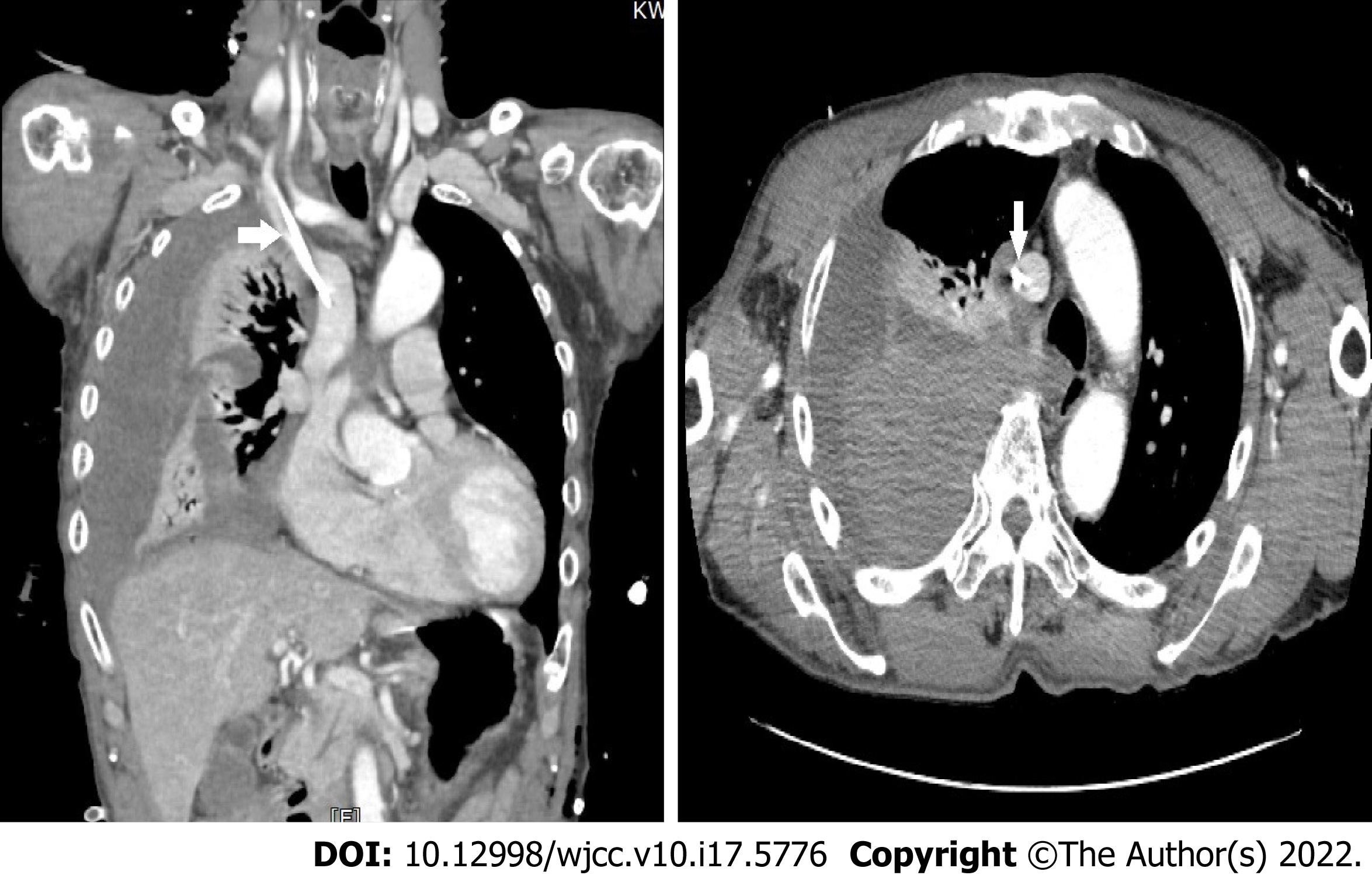
Figure 2 Postoperative chest computed tomography taken upon arrival in the recovery room.
There is large amount of hemothorax in the right hemithorax with no evidence of extravasation of contrast media. The central catheter (arrows) is located within the superior vena cava
- Citation: Kang H, Cho SY, Suk EH, Ju W, Choi JY. Massive hemothorax following internal jugular vein catheterization under ultrasound guidance: A case report. World J Clin Cases 2022; 10(17): 5776-5782
- URL: https://www.wjgnet.com/2307-8960/full/v10/i17/5776.htm
- DOI: https://dx.doi.org/10.12998/wjcc.v10.i17.5776