Copyright
©The Author(s) 2022.
World J Clin Cases. Jun 6, 2022; 10(16): 5124-5132
Published online Jun 6, 2022. doi: 10.12998/wjcc.v10.i16.5124
Published online Jun 6, 2022. doi: 10.12998/wjcc.v10.i16.5124
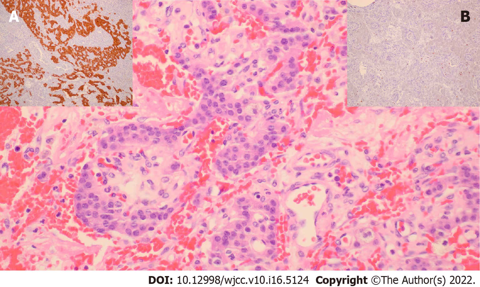
Figure 2 Pathohistological appearance of malignant insulinoma.
One of our own cases, G2 pancreatic neuroendocrine tumor, exhibited small nests of uniform cells showing infiltrative growth pattern (hemalaun and eosin, × 400). A; Brown cytoplasmic staining in tumorous cells (synaptophysin, × 100), B; Low Ki-67 proliferation index of up to 7% (Ki-67, ×200).
- Citation: Cigrovski Berkovic M, Ulamec M, Marinovic S, Balen I, Mrzljak A. Malignant insulinoma: Can we predict the long-term outcomes? World J Clin Cases 2022; 10(16): 5124-5132
- URL: https://www.wjgnet.com/2307-8960/full/v10/i16/5124.htm
- DOI: https://dx.doi.org/10.12998/wjcc.v10.i16.5124


