Copyright
©The Author(s) 2022.
World J Clin Cases. May 16, 2022; 10(14): 4586-4593
Published online May 16, 2022. doi: 10.12998/wjcc.v10.i14.4586
Published online May 16, 2022. doi: 10.12998/wjcc.v10.i14.4586
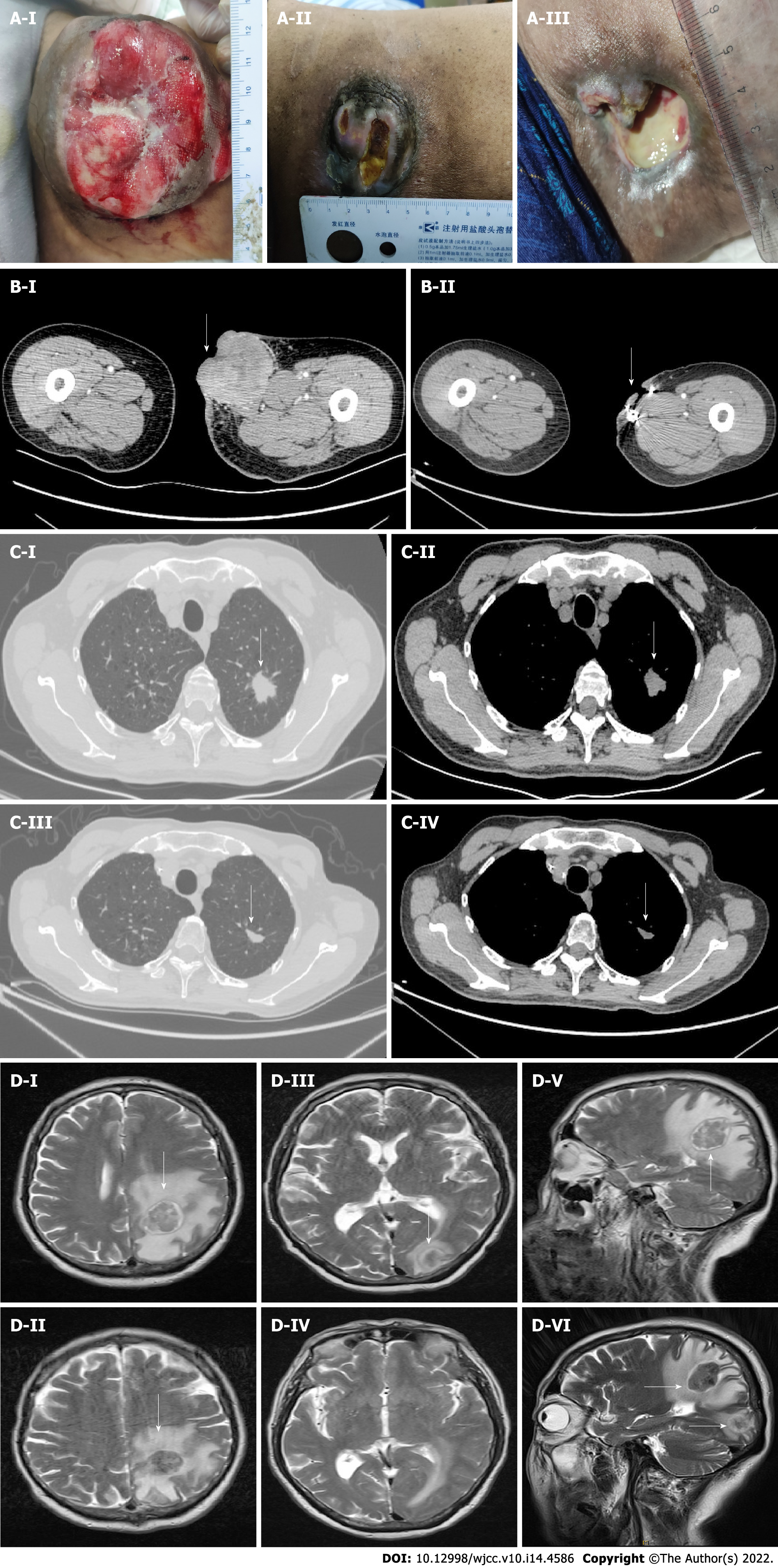
Figure 2 Comparison of lesion size before the initial stage treatment and after 6 cycles of maintenance treatment.
A: Lesion size before treatment (A-I). Lesion size after 6 cycles of the initial stage chemotherapy (A-II). Lesion size after 6 cycles of maintenance treatment (A-III); B: CT scans showing comparison of lesion size in the thigh. Before treatment = about 68 mm × 74 mm (B-I), after 6 cycles of maintenance treatment = about 36 mm × 14 mm (B-II); C: CT scans showing comparison of lesion size in the lung. Before treatment = 23 mm × 21 mm × 25 mm (C-I and C-II), after treatment = 11 mm × 17 mm × 13 mm (C-III and C-IV); D: MRI results showing comparison of lesion size in the brain. Lesion in the left parietal lobe before treatment = 32 mm × 28 mm × 29 mm (D-I, D-III and D-V), after treatment, lesion in the left parietal lobe = 25 mm × 22 mm, new lesion in the left occipital lobe = 18 mm × 15 mm (D-II, D-IV and D-VI) (white arrow).
- Citation: Wei XL, Liu Q, Zeng QL, Zhou H. Primary or metastatic lung cancer? Sebaceous carcinoma of the thigh: A case report. World J Clin Cases 2022; 10(14): 4586-4593
- URL: https://www.wjgnet.com/2307-8960/full/v10/i14/4586.htm
- DOI: https://dx.doi.org/10.12998/wjcc.v10.i14.4586


