Copyright
©The Author(s) 2022.
World J Clin Cases. May 6, 2022; 10(13): 4301-4313
Published online May 6, 2022. doi: 10.12998/wjcc.v10.i13.4301
Published online May 6, 2022. doi: 10.12998/wjcc.v10.i13.4301
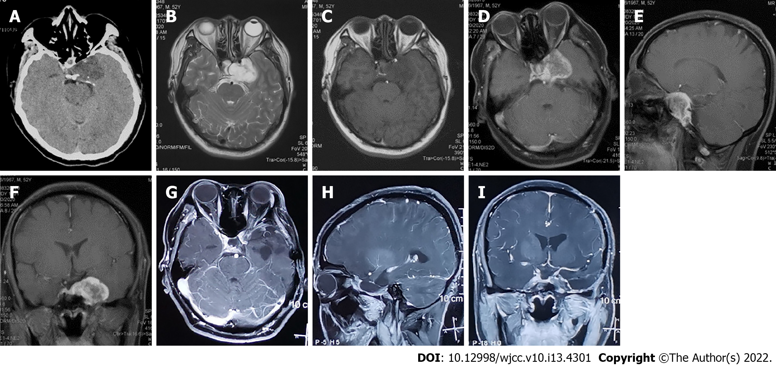
Figure 1 Preoperative and Postoperative follow-up imaging examination results.
A: Computed tomography scan of the patient’s head at admission. B-F: Preoperative brain magnetic resonance imaging (MRI), B: axial view of T2-weighted image; C: axial view of T1-weighted image. D-F: Axial, sagittal and coronal view of gadolinium-injected enhancement MRI scan. G-I: Axial, sagittal, coronal view of postoperative brain gadolinium-enhanced MRI scan in 12 mo after surgery.
- Citation: Zhu ZY, Wang YB, Li HY, Wu XM. Primary intracranial extraskeletal myxoid chondrosarcoma: A case report and review of literature. World J Clin Cases 2022; 10(13): 4301-4313
- URL: https://www.wjgnet.com/2307-8960/full/v10/i13/4301.htm
- DOI: https://dx.doi.org/10.12998/wjcc.v10.i13.4301


