Copyright
©The Author(s) 2022.
World J Clin Cases. Jan 7, 2022; 10(1): 143-154
Published online Jan 7, 2022. doi: 10.12998/wjcc.v10.i1.143
Published online Jan 7, 2022. doi: 10.12998/wjcc.v10.i1.143
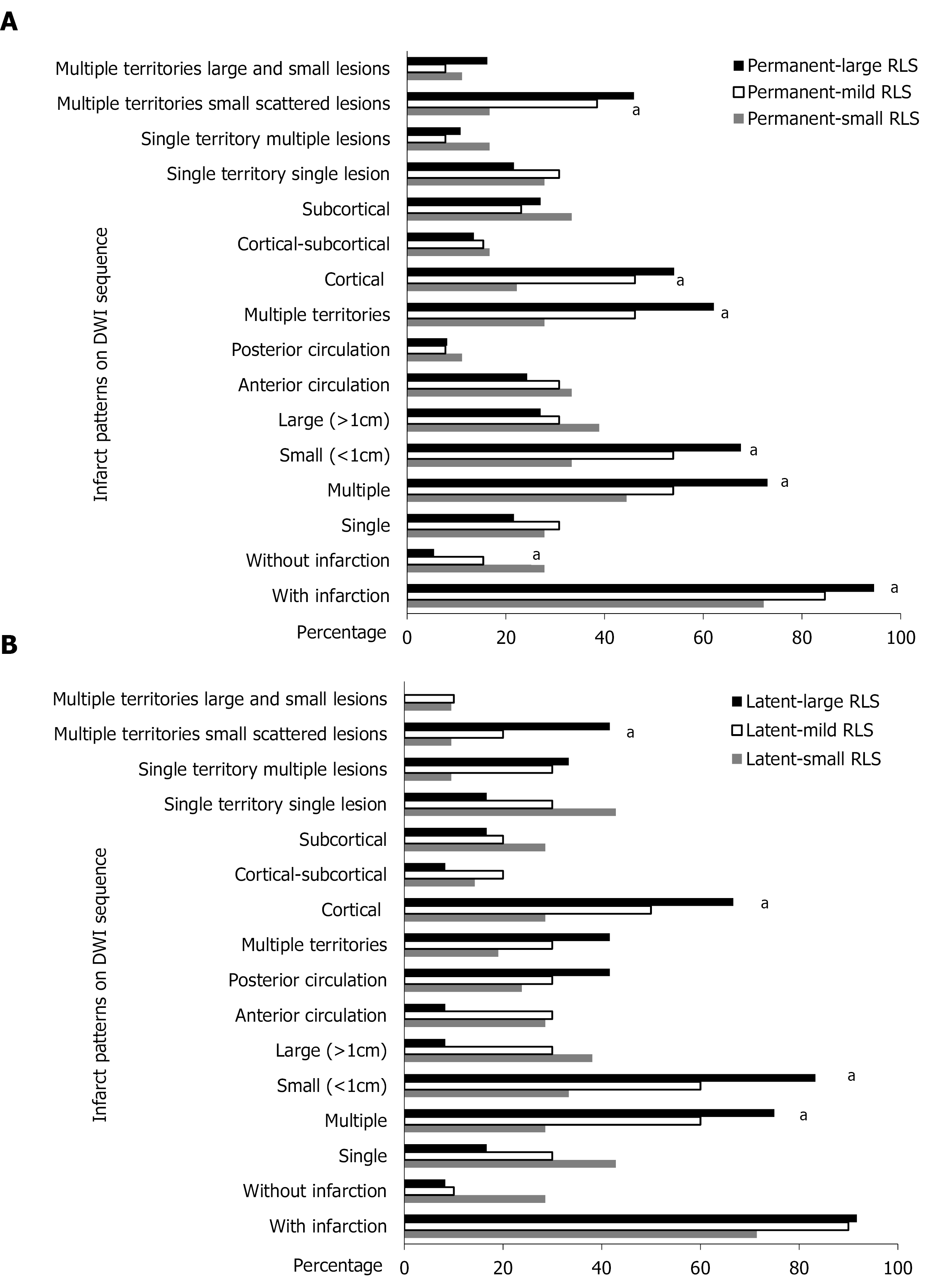
Figure 4 Diffusion-weighted imaging lesions of different grades of shunt in permanent and latent right-to-left shunt groups.
A: Infarction pattern of permanent-small, permanent-mild and permanent-large right-to-left shunt (RLS). Multiple, small and cortical lesions were more likely to be involved as the grade of shunt increased in the permanent RLS group; B: Infarction pattern of latent-small, latent-mild and latent-large RLS. Multiple, small and cortical lesions were more likely to be involved as the grade of shunt increased in the latent RLS group. aP < 0.05.
- Citation: Xiao L, Yan YH, Ding YF, Liu M, Kong LJ, Hu CH, Hui PJ. Evaluation of right-to-left shunt on contrast-enhanced transcranial Doppler in patent foramen ovale-related cryptogenic stroke: Research based on imaging. World J Clin Cases 2022; 10(1): 143-154
- URL: https://www.wjgnet.com/2307-8960/full/v10/i1/143.htm
- DOI: https://dx.doi.org/10.12998/wjcc.v10.i1.143


