Copyright
©The Author(s) 2020.
World J Clin Cases. Nov 6, 2020; 8(21): 5426-5431
Published online Nov 6, 2020. doi: 10.12998/wjcc.v8.i21.5426
Published online Nov 6, 2020. doi: 10.12998/wjcc.v8.i21.5426
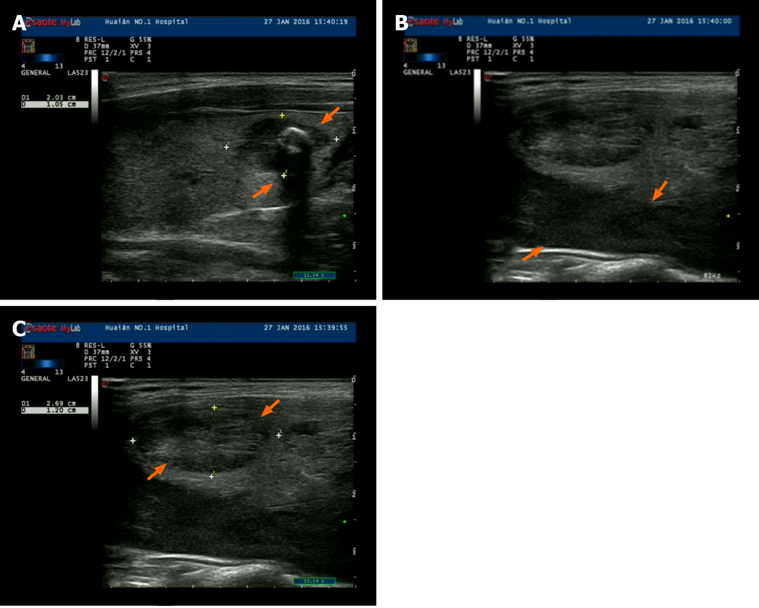
Figure 1 Ultrasound images of the patient.
A: Papillary thyroid carcinoma; B: Parathyroid adenoma; C: Thyroid adenoma.
Figure 2 Magnetic resonance imaging of the right inferior parathyroid (orange arrows).
A: Coronal section; B: Transverse section; C: Sagittal section.
Figure 3 Histology of parathyroid adenoma, papillary thyroid carcinoma and thyroid adenoma.
A: Parathyroid adenoma [hematoxylin and eosin (H&E) staining, 400 ×, insert 1000 ×]; B: Papillary thyroid carcinoma (H&E staining, 400 ×, insert 1000 ×); C: Thyroid follicular adenomas (H&E staining, 100 ×, insert 400 ×).
- Citation: Li Q, Xu XZ, Shi JH. Synchronous parathyroid adenoma, papillary thyroid carcinoma and thyroid adenoma in pregnancy: A case report. World J Clin Cases 2020; 8(21): 5426-5431
- URL: https://www.wjgnet.com/2307-8960/full/v8/i21/5426.htm
- DOI: https://dx.doi.org/10.12998/wjcc.v8.i21.5426


