Copyright
©The Author(s) 2020.
World J Clin Cases. Nov 6, 2020; 8(21): 5070-5085
Published online Nov 6, 2020. doi: 10.12998/wjcc.v8.i21.5070
Published online Nov 6, 2020. doi: 10.12998/wjcc.v8.i21.5070
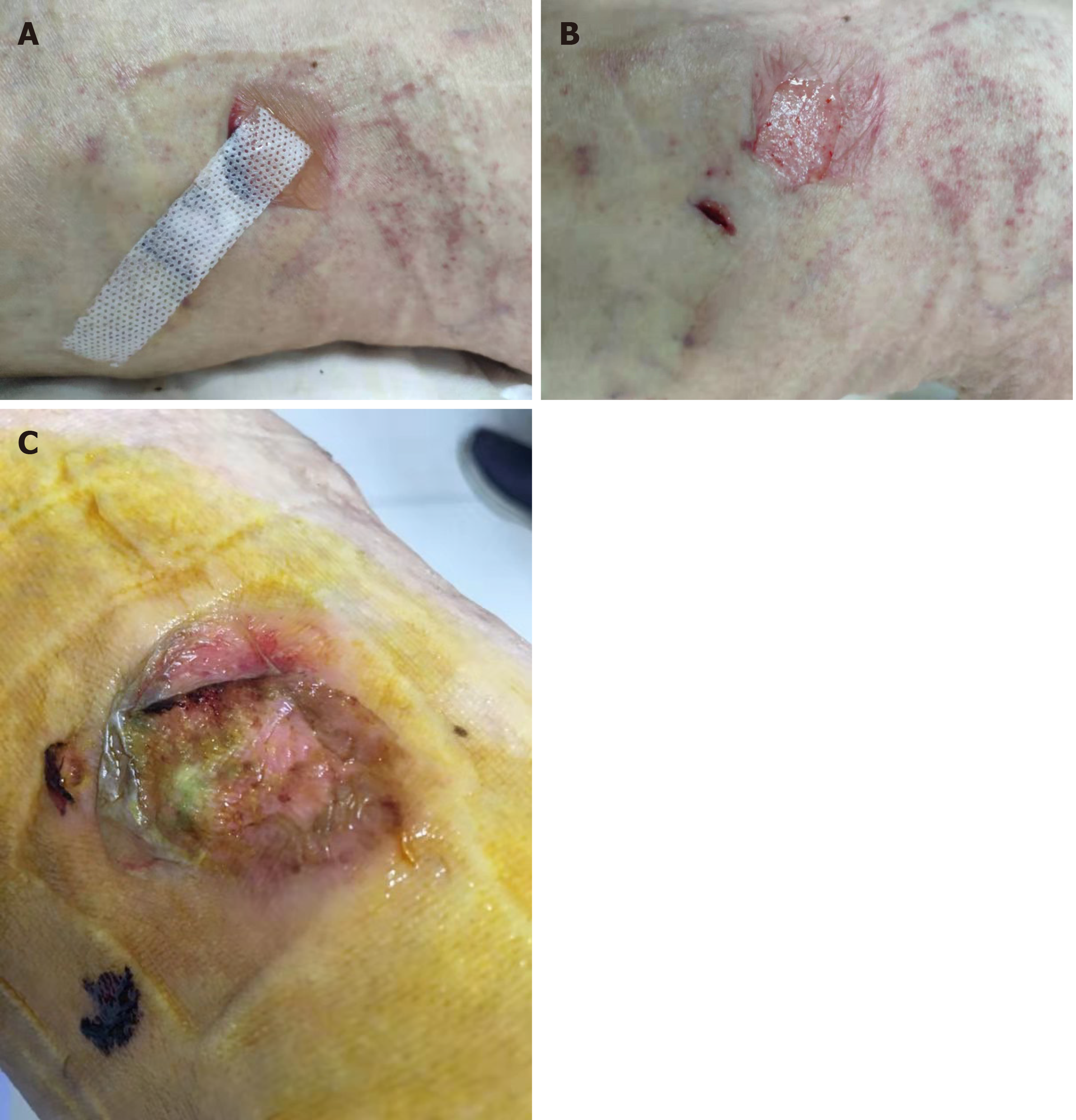
Figure 1 Patient received minimally invasive surgery for varicose veins, the small incision was closed by adhesive strap on November 4, 2019.
A: An allergic blister was found on postprocedure day 4; B: The skin was peeled while removing the strap; C: The wound was unhealed on November 12, 2019 as the patient failed to follow-up on schedule for wound care.
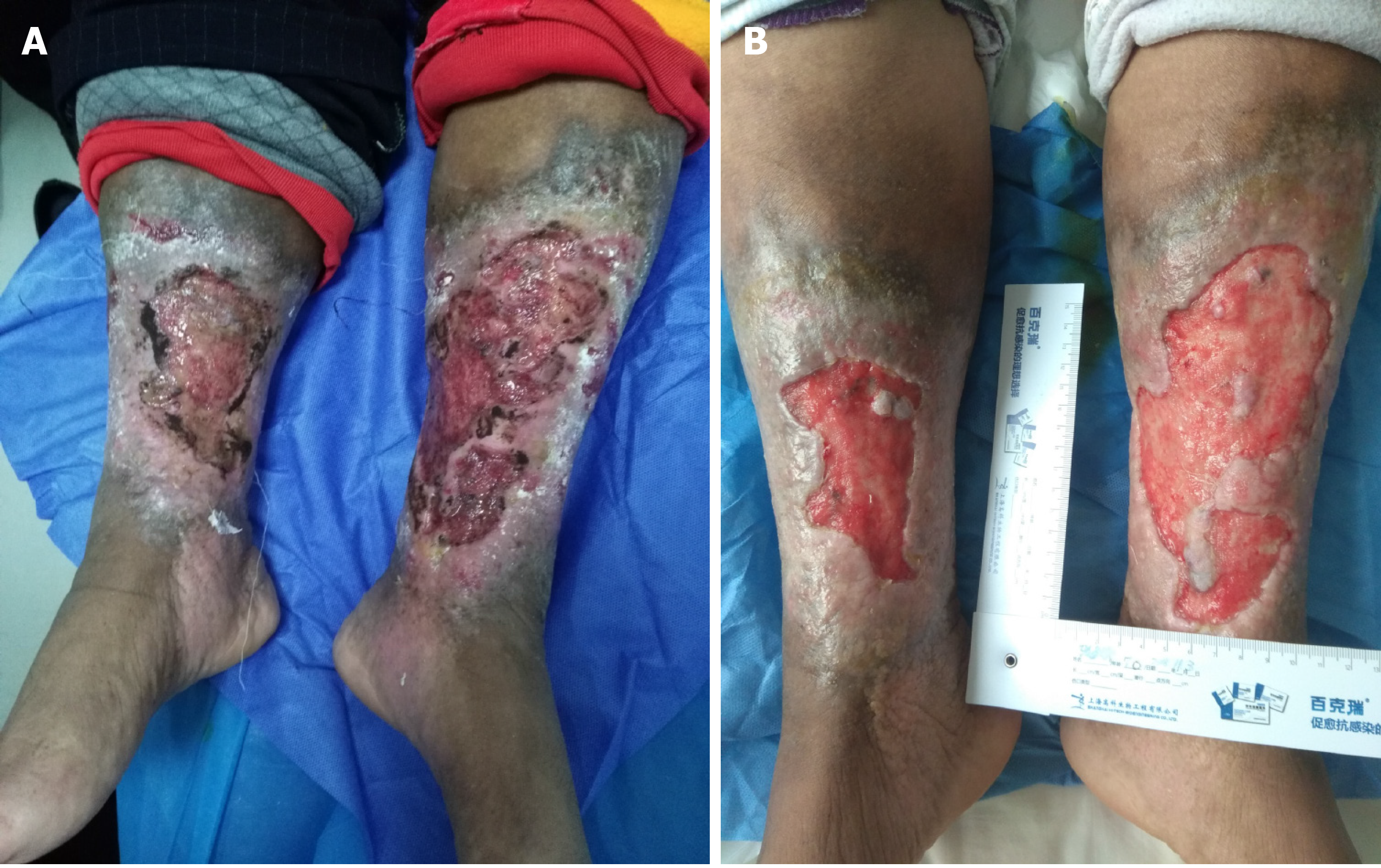
Figure 2 This 70-year-old female patient had suffered from chronic venous leg ulcers on both lower extremities for 40 years.
Thirty years ago, a skin graft was harvested from her thigh without any anesthesia expecting for better survival of the graft. Unfortunately, the skin graft failed to grow, and she refused any skin harvest for skin graft. At this current time, her wound was prepared well enough for skin graft. A: Chronic venous leg ulcers on both lower extremities; B: Prepared for skin graft.
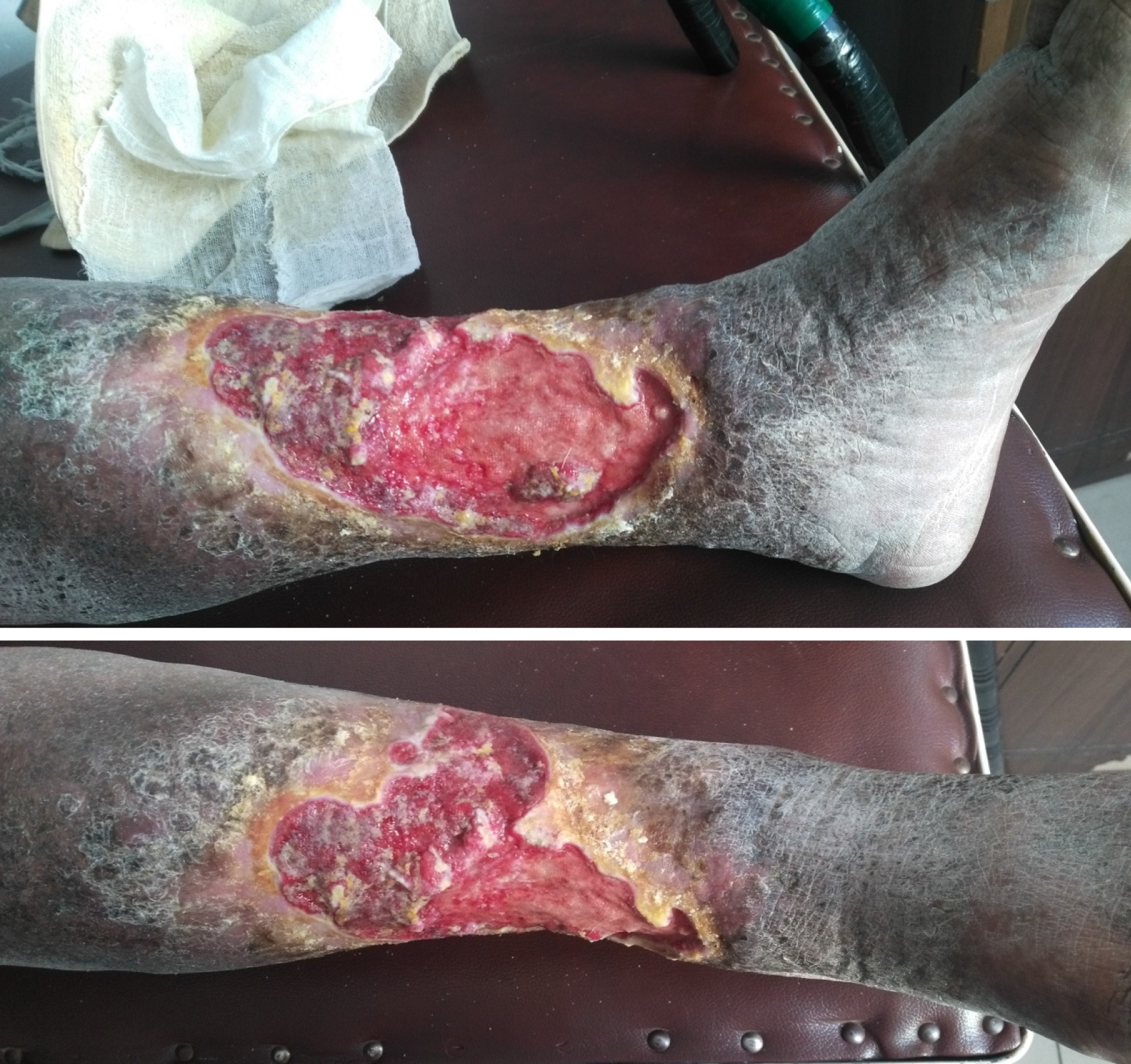
Figure 3 The patient had chronic venous leg ulcers for approximately 20 years.
The pathological study of a biopsy from the wound indicated squamous cell carcinoma.
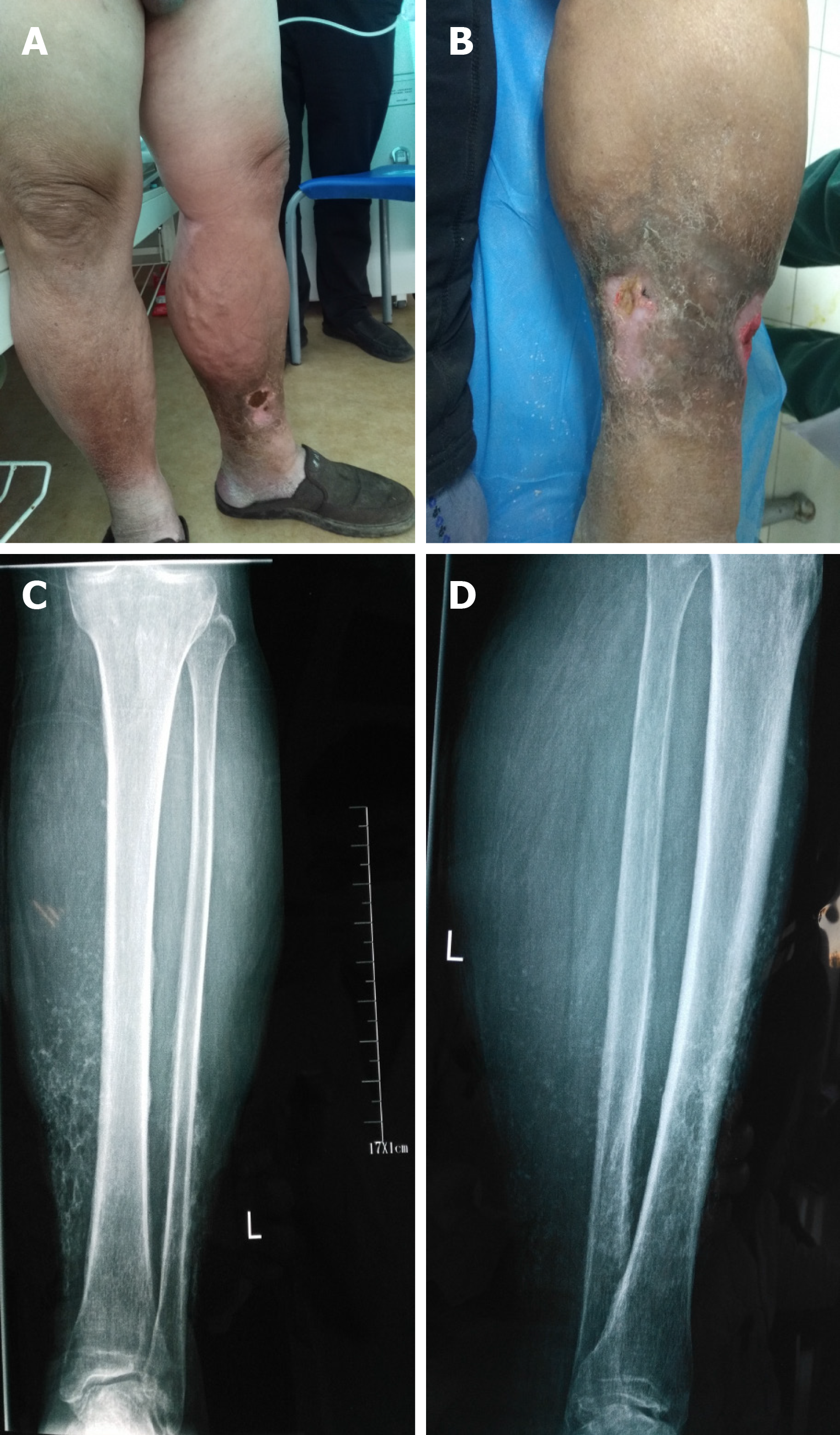
Figure 4 The male patient had varicose vein and venous insufficiency for 15 years and ulcers for 1.
5 years. A: Redness, warmth, pain, and edema with two ulcers were noticed on admission; B: The inflation of the skin was relived after initial treatment; C and D: X-ray showed calcification of the tissue around ulcers.
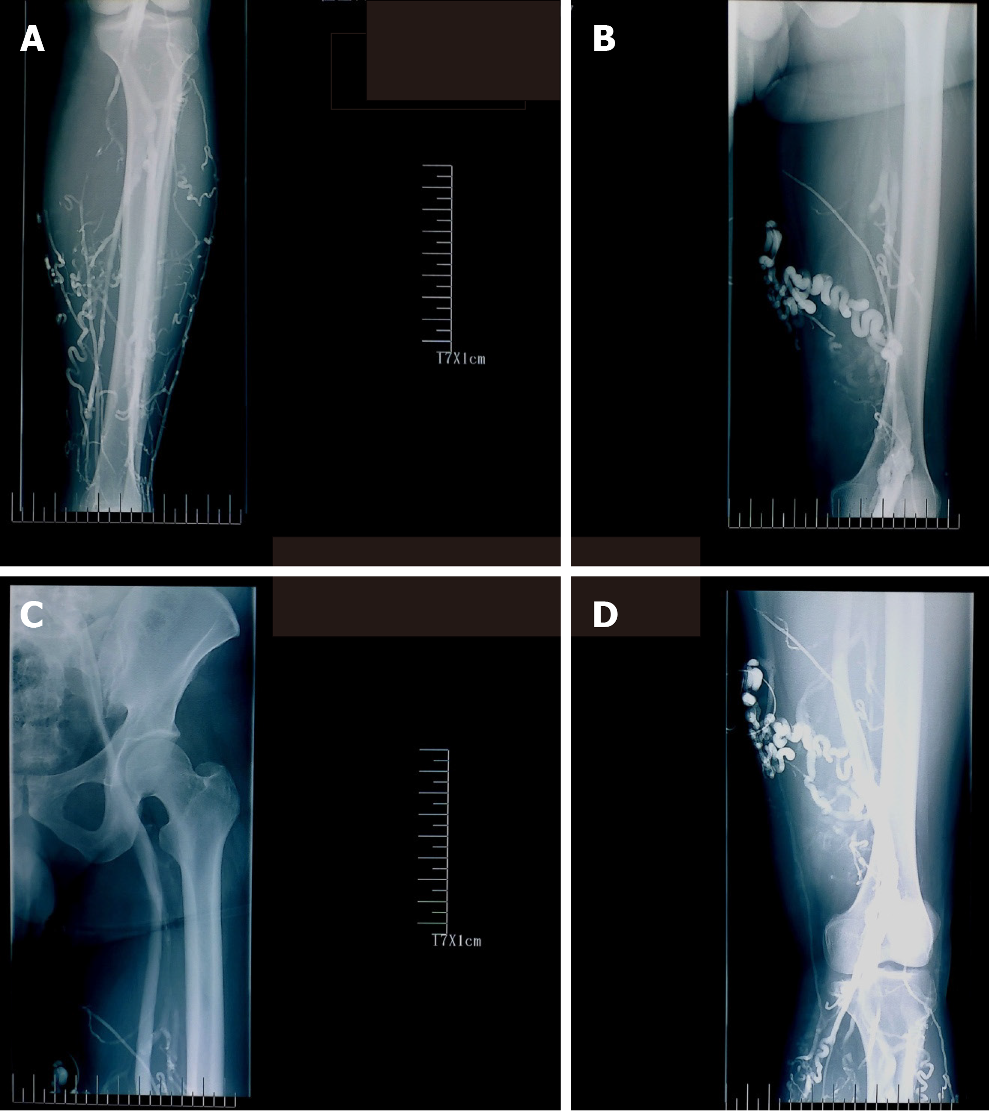
Figure 5 Ascending venogram shows the varicosities at lower leg and thigh, patency of femoral and iliac veins, and the degree of reflux while performing venography.
A: Lower leg; B and D: Thigh; C: Femoral and iliac veins.
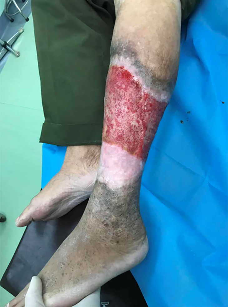
Figure 6 The edge of the ulcer appears white, a sign for the development of granulation tissue.
This tissue should be preserved rather removed during debridement.
- Citation: Ren SY, Liu YS, Zhu GJ, Liu M, Shi SH, Ren XD, Hao YG, Gao RD. Strategies and challenges in the treatment of chronic venous leg ulcers. World J Clin Cases 2020; 8(21): 5070-5085
- URL: https://www.wjgnet.com/2307-8960/full/v8/i21/5070.htm
- DOI: https://dx.doi.org/10.12998/wjcc.v8.i21.5070


