Copyright
©The Author(s) 2019.
World J Clin Cases. Jan 26, 2019; 7(2): 253-259
Published online Jan 26, 2019. doi: 10.12998/wjcc.v7.i2.253
Published online Jan 26, 2019. doi: 10.12998/wjcc.v7.i2.253
Figure 1 Preoperative body examination and magnetic resonance imaging.
A: A T-tube for biliary drainage with grey-white, hyperplastic granulation tissue (with a stench) surrounding the tube; B: Magnetic resonance imaging demonstrates a huge mass in the gallbladder area, embedding in the common bile duct where a drainage tube was seen.
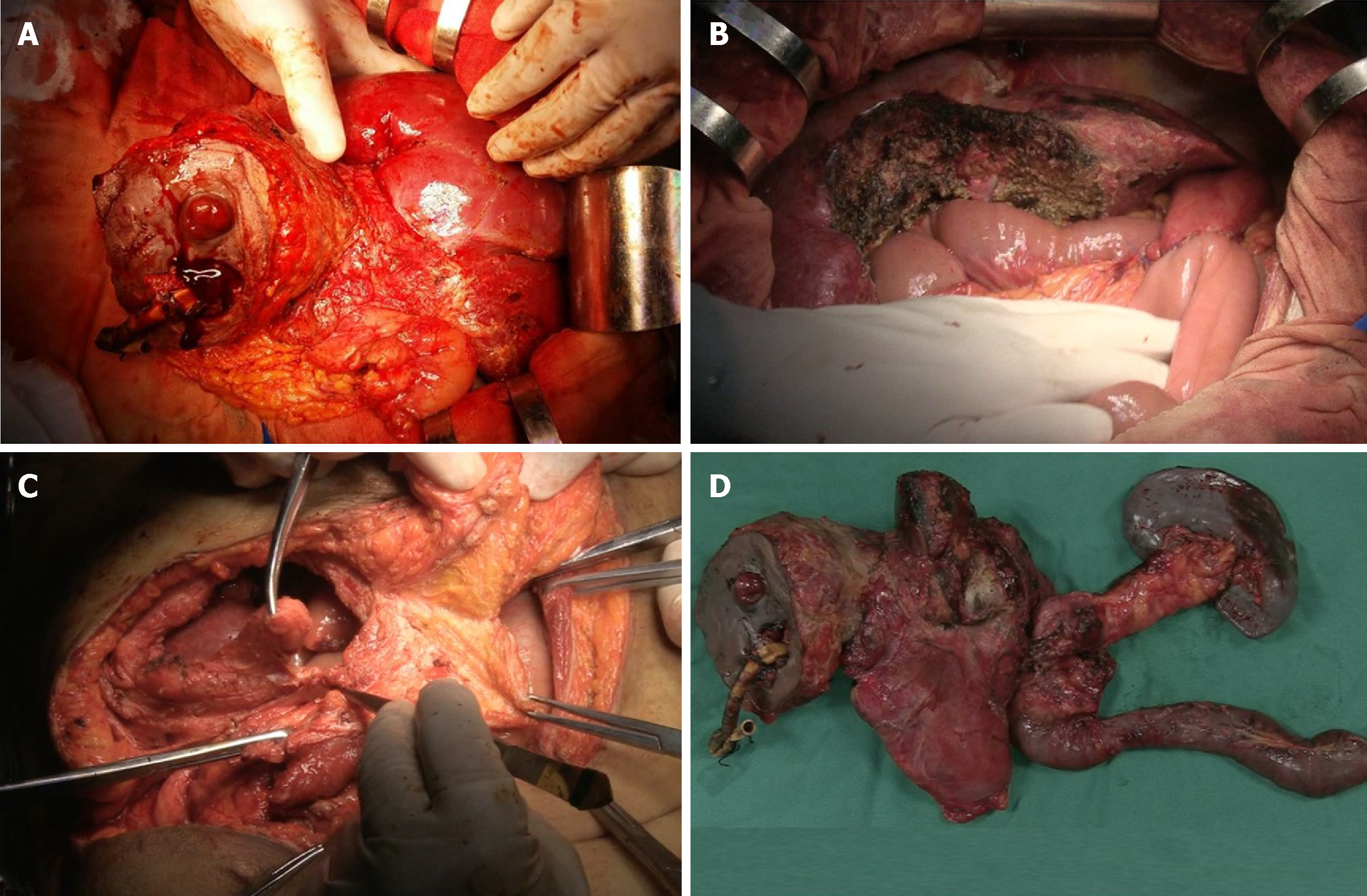
Figure 2 Surgical procedure.
A: Dissecting the abdominal wall around the T-tube step by step; B: Choledochojejunostomy, gastrointestinal anastomosis, and intestinal anastomosis were performed after removing all lesions; C: Closing the incision by transferring rectus abdominis and external oblique muscle flap; D: The en bloc specimen of the liver, common bile duct, duodenum, partial stomach, spleen, and pancreas was removed.
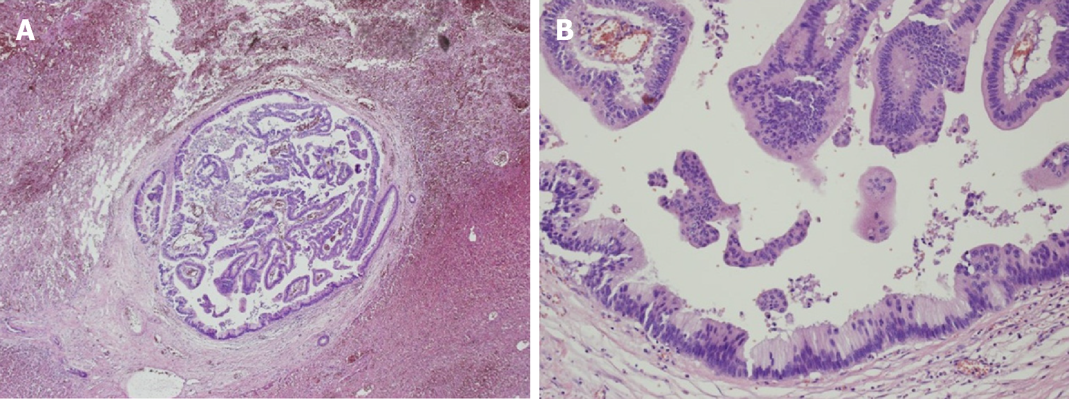
Figure 3 Histological examination (magnification, × 400) revealed a moderately differentiated adenocarcinoma originating from the biliary system.
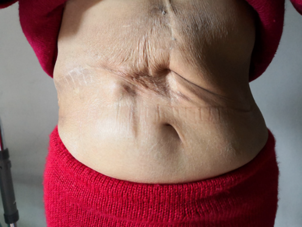
Figure 4 Surgical incision at 35 mo after operation.
She was asymptomatic and in good physical condition.
- Citation: Xiao Y, Zhao J, Wu H, Xie KL, Wan Y, Xu XW, Zhang YG. Surgical treatment of malignant biliary papillomatosis invading adjacent organs: A case report. World J Clin Cases 2019; 7(2): 253-259
- URL: https://www.wjgnet.com/2307-8960/full/v7/i2/253.htm
- DOI: https://dx.doi.org/10.12998/wjcc.v7.i2.253


