Copyright
©The Author(s) 2019.
World J Clin Cases. Jan 26, 2019; 7(2): 221-227
Published online Jan 26, 2019. doi: 10.12998/wjcc.v7.i2.221
Published online Jan 26, 2019. doi: 10.12998/wjcc.v7.i2.221
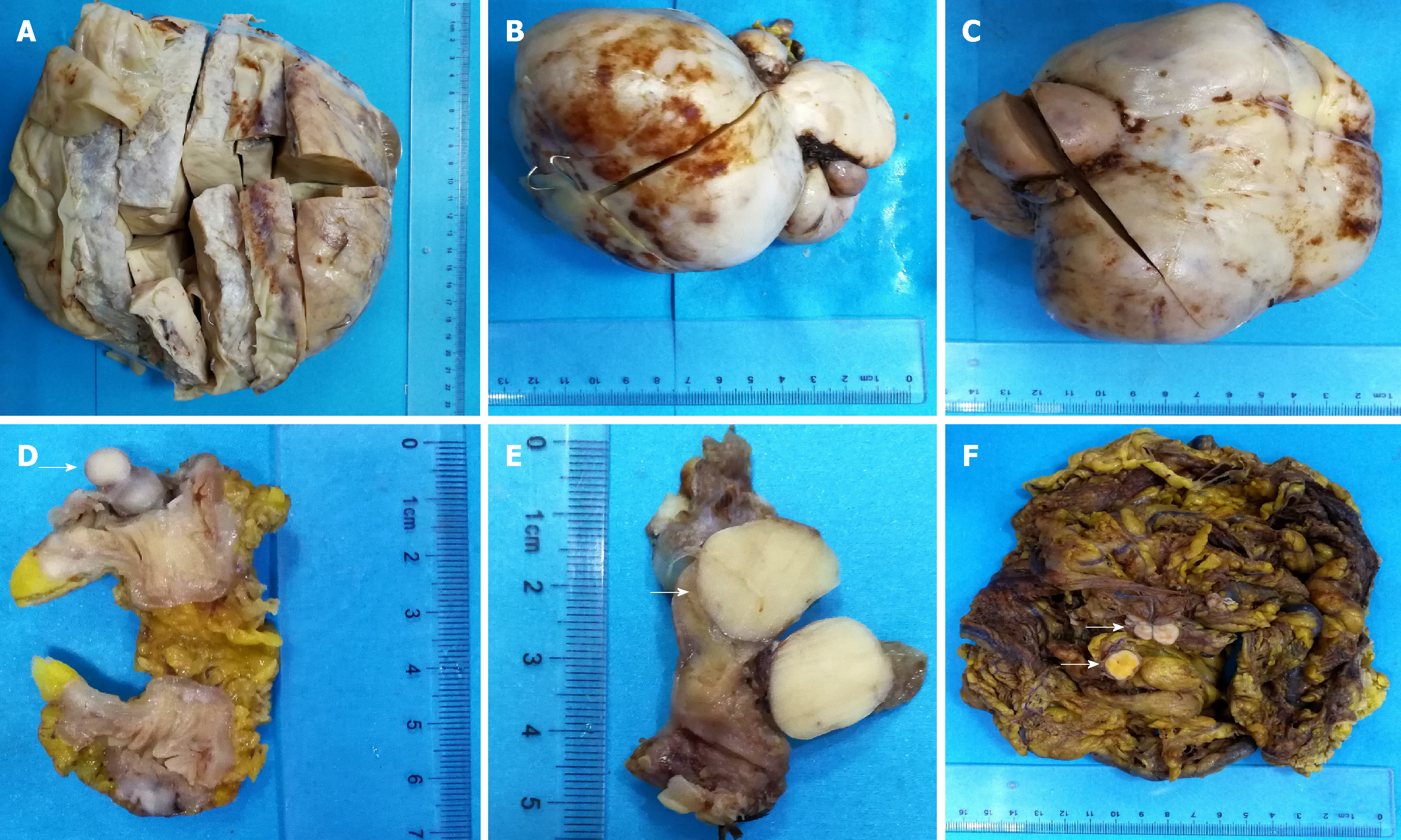
Figure 1 General view of low-grade endometrial stromal sarcoma.
A: A pelvic mass with a maximum diameter of 25 cm; B: A mass from the abdominal cavity with a maximum diameter of 13 cm; C: A mass from the abdominal cavity with a maximum diameter of 15 cm; D: A mass from the small intestine wall with a maximum diameter of 1.5 cm (arrow); E: A mass from the uterine tube wall with a maximum diameter of 1.5 cm (arrow); F: Omentum tissues, in which several scattered nodules were shown with diameters of 1.5-2.5 cm (arrows).
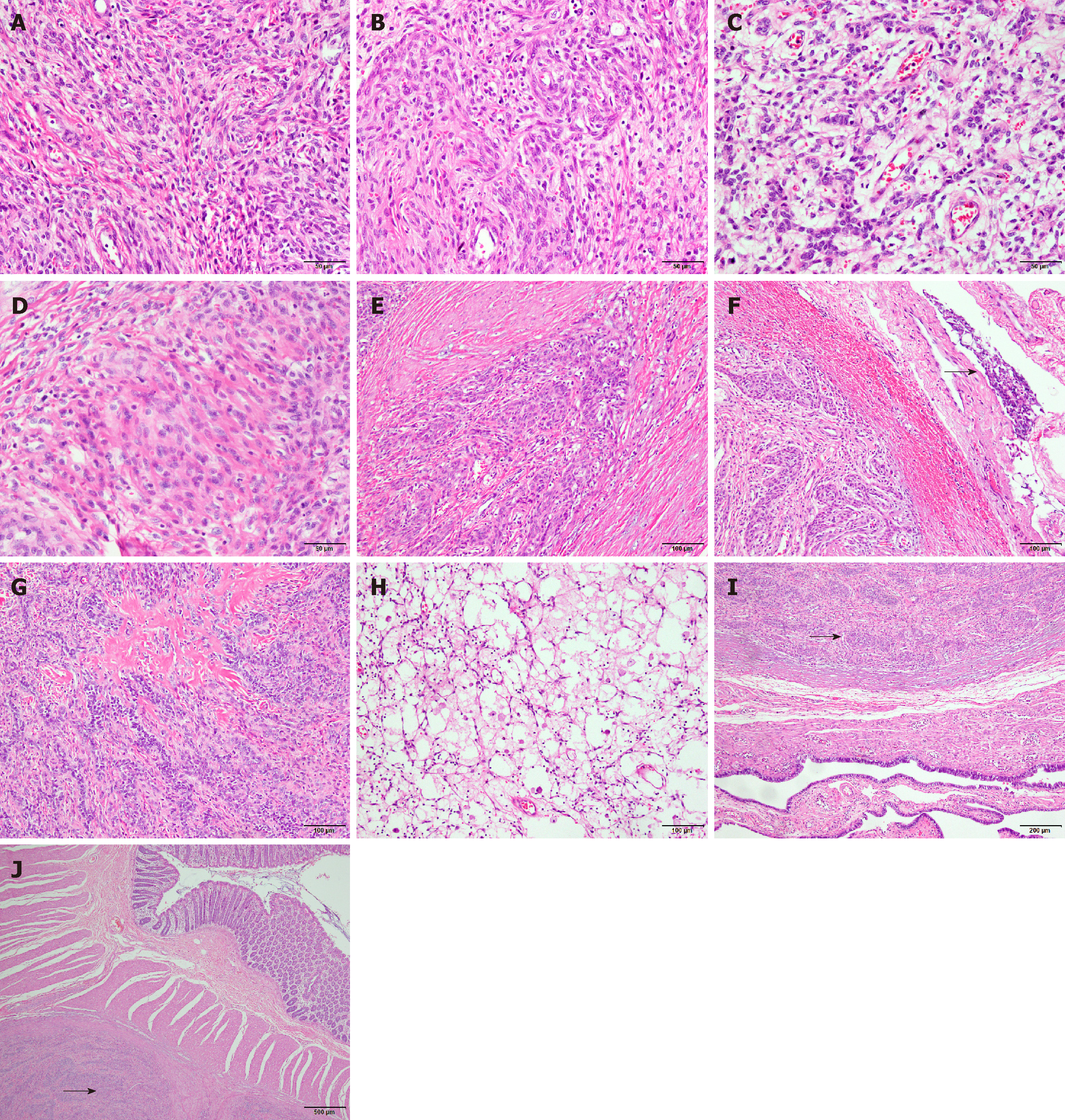
Figure 2 Primary microscopic morphological characteristics of low-grade endometrial stromal sarcoma.
A: Tumor cells are diffusely distributed, uniformly sized, and short spindle-shaped; the cells are benign, and mitotic figures are rare (original magnification, ×400); B: Tumor cells are swirled around the blood vessels (original magnification, ×400); C: Sex cord-like differentiated tumor cells arranged in cords (original magnification, ×400); D: Smooth muscle-like differentiated tumor cells (original magnification, ×400); E: Tumor cells showing tongue-like invasive growth (original magnification, ×400); F: Tumor cells invading the encapsulated vessel, indicated by the arrow (original magnification, ×200); G: Stroma showing obvious hyaline degeneration, forming a starburst-like structure (original magnification, ×200); H: Many foam cells and inflammatory cells are visible in some localized areas (original magnification, ×200); I: The tumor invading the muscle wall of the oviduct, indicated by the arrow (original magnification, ×100); J: The tumor invading the wall of the small intestine, indicated by the arrow (original magnification, ×40).
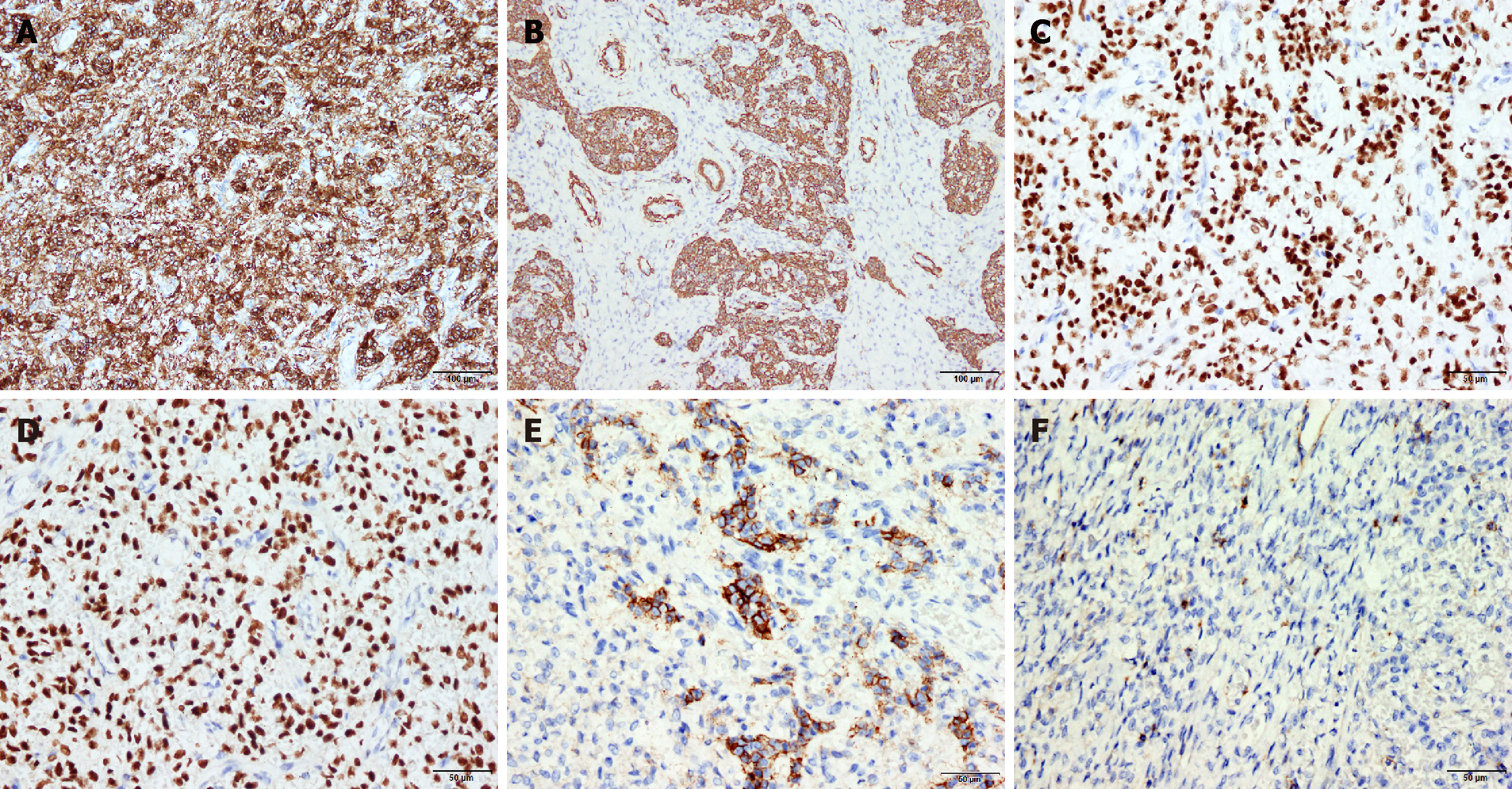
Figure 3 Main immunohistochemical staining results for low-grade endometrial stromal sarcoma.
A: Tumor cells are strongly diffusely positive for CD10 (original magnification, ×200); B: Tumor cells are partially positive for SMA (original magnification, ×200); C: Tumor cells are diffusely positive for ER (>90%) (original magnification, ×400); D: Tumor cells are diffusely positive for PR (>90%) (original magnification, ×400); E: Tumor cells are partially positive for CD56 (original magnification, ×400); F: Tumor cells are partially positive for CD99 (original magnification, ×400).
- Citation: Zhu Q, Sun YQ, Di XQ, Huang B, Huang J. Metastatic low-grade endometrial stromal sarcoma with sex cord and smooth muscle differentiation: A case report. World J Clin Cases 2019; 7(2): 221-227
- URL: https://www.wjgnet.com/2307-8960/full/v7/i2/221.htm
- DOI: https://dx.doi.org/10.12998/wjcc.v7.i2.221


