Copyright
©The Author(s) 2017.
World J Clin Cases. Jul 16, 2017; 5(7): 258-263
Published online Jul 16, 2017. doi: 10.12998/wjcc.v5.i7.258
Published online Jul 16, 2017. doi: 10.12998/wjcc.v5.i7.258
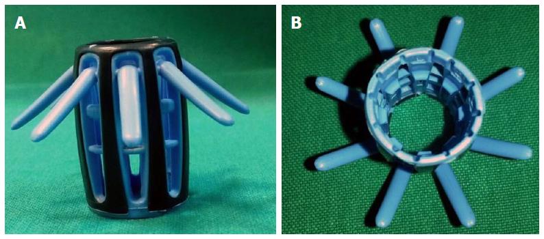
Figure 1 Endocuff’s view: (A) lateral; (B) from above.
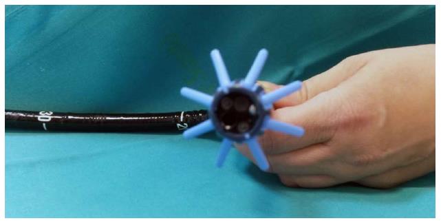
Figure 2 Endocuff Vision™ mounted at the tip of the colonoscope.
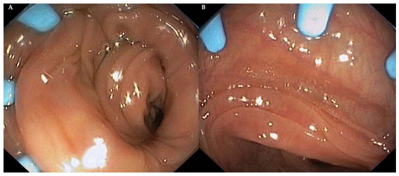
Figure 3 Endoscopic view of colonoscope retraction in which it’s possible to note the flatting folds.
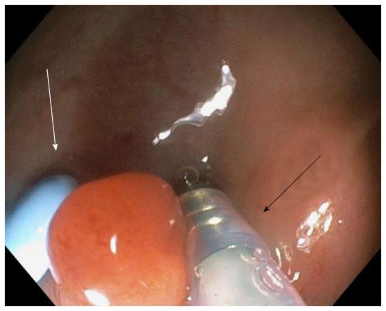
Figure 4 Endoscopic removal of a sessile polyp of the sigmoid colon, in which it’s possible to see the Endocuff’s flat (white arrow) and injection needle (22 G, Micro-Tech, Nanjing Co, Ltd) (black arrow).
- Citation: Zippi M, Hong W, Crispino P, Traversa G. New device to implement the adenoma detection rate. World J Clin Cases 2017; 5(7): 258-263
- URL: https://www.wjgnet.com/2307-8960/full/v5/i7/258.htm
- DOI: https://dx.doi.org/10.12998/wjcc.v5.i7.258


