Copyright
©The Author(s) 2015.
World J Clin Cases. May 16, 2015; 3(5): 442-449
Published online May 16, 2015. doi: 10.12998/wjcc.v3.i5.442
Published online May 16, 2015. doi: 10.12998/wjcc.v3.i5.442
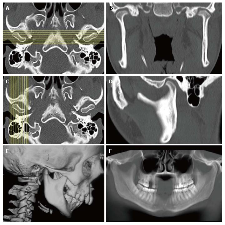
Figure 1 Technique of reconstruction.
A, B: Sagittal images are reconstructed perpendicular to the plane of the glenoid fossa; C, D: Coronal images are reconstructed perpendicular to the plane of sagittal images; E, F: The joint anatomy is well depicted in 3D reformatted and reconstructed panoramic image.
Figure 2 Goldenhaar Syndrome in a young boy.
Shaded surface display (A) image shows micrognathia, downward slanting left palpebral fissure and hypoplastic left pinna. 3D reformatted images show the normal right hemimandible (B) and hypoplastic body and ramus of left hemimandible with underdeveloped mandibular condyle and glenoid fossa (C).
Figure 3 Mandibular hypoplasia in a 7-year-old boy.
Axial (A), coronal (B) and 3D reformatted (C) images show show small left hemi-mandible with hypoplastic mandibular condyle and glenoid fossa (arrows).
Figure 4 Condylar hyperplasia.
Axial (A), coronal (B) and 3D reformatted images (C) show enlarged left mandibular ramus and condyle (arrows).
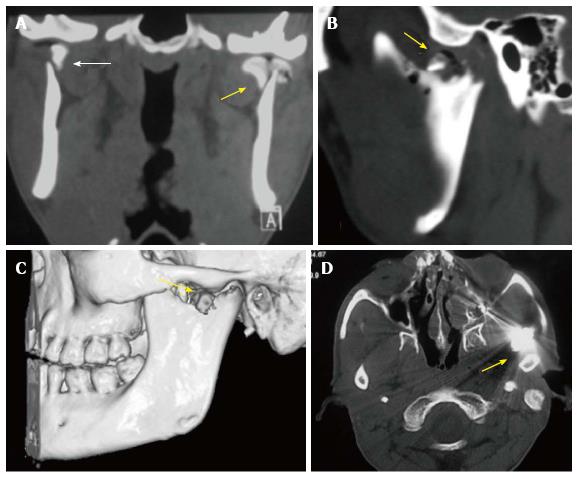
Figure 5 Condylar fractures in different patients.
Coronal (A), sagittal (B) and 3D (C) reconstructed CT images depict bilateral displaced, extracapsular (right- bold arrow), and intracapsular (left- arrow) condylar head fractures with involvement of the articular surface on the left side. The intra-articular air on the left side indicates an open wound (arrow). Open joint wound with a foreign body (bullet in another patient- D) or gross contamination are indications for open surgery.
Figure 6 Pyogenic arthritis in a 33-year-old woman with fever and pain around right ear.
A: Soft tissue edema with loss of fat planes (arrow) around right temporomandibular joint is seen in axial computed tomography (CT) image (A); B: Bony erosions in the condyle are well seen in axial CT image in bone window setting (arrow).
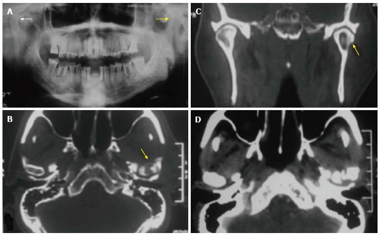
Figure 7 Chronic osteomyelitis of temporomandibular joint in a 55-year-old patient.
Sclerosis of left mandibular condyle is seen in panorex radiograph (A); a bony sequestrum is seen within the left mandibular condyle in axial (B) and coronal (C) reformatted computed tomography (CT) images; axial CT section in soft tissue window (D) shows inflammatory changes in tissues around the temporomandibular joint.
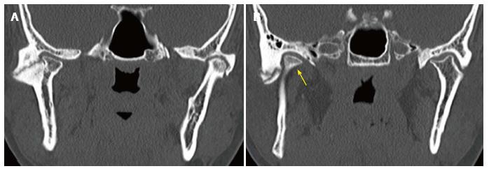
Figure 8 Post traumatic ankylosis in a 28-year-old male patient.
Coronal reformatted multi-detector computed tomography images demonstrate ankylosis and pseudo-joint formation in both temporomandibular joints (A); mandibular condyle is displaced anteriorly and medially (B). The referring clinician needs to know the relationship of the condylar mass to adjacent skull base structures.
Figure 9 Osteochondroma in a 32-year-old man presenting with trismus.
Sagittal (A) and 3D reconstructed (B) images demonstrate a bony outgrowth arising from the mandibular condyle (arrow), just beneath the articular eminence.
Figure 10 Jacob’s disease in a 17-year-old boy presenting with restricted mouth opening.
Axial (A), sagittal (B) reformatted computed tomography images depict “pseudo-joint formation” between enlarged coronoid process of mandible and zygomatic arch (arrows).
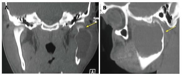
Figure 11 Aneurysmal bone cyst of the mandible in a 17-year-old boy present with left cheek swelling since one year.
Coronal (A) and sagittal (B) reformatted computed tomography images depict a large, expansile, lytic lesion involving left mandibular condyle and ramus.
- Citation: Pahwa S, Bhalla AS, Roychaudhary A, Bhutia O. Multidetector computed tomography of temporomandibular joint: A road less travelled. World J Clin Cases 2015; 3(5): 442-449
- URL: https://www.wjgnet.com/2307-8960/full/v3/i5/442.htm
- DOI: https://dx.doi.org/10.12998/wjcc.v3.i5.442


