
















©The Author(s) 2015.
World J Clin Cases. May 16, 2015; 3(5): 442-449
Published online May 16, 2015. doi: 10.12998/wjcc.v3.i5.442
Published online May 16, 2015. doi: 10.12998/wjcc.v3.i5.442
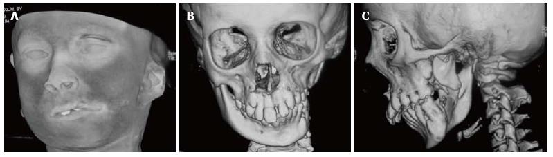
Figure 2 Goldenhaar Syndrome in a young boy.
Shaded surface display (A) image shows micrognathia, downward slanting left palpebral fissure and hypoplastic left pinna. 3D reformatted images show the normal right hemimandible (B) and hypoplastic body and ramus of left hemimandible with underdeveloped mandibular condyle and glenoid fossa (C).
- Citation: Pahwa S, Bhalla AS, Roychaudhary A, Bhutia O. Multidetector computed tomography of temporomandibular joint: A road less travelled. World J Clin Cases 2015; 3(5): 442-449
- URL: https://www.wjgnet.com/2307-8960/full/v3/i5/442.htm
- DOI: https://dx.doi.org/10.12998/wjcc.v3.i5.442