Copyright
©The Author(s) 2023.
World J Clin Cases. Apr 16, 2023; 11(11): 2423-2434
Published online Apr 16, 2023. doi: 10.12998/wjcc.v11.i11.2423
Published online Apr 16, 2023. doi: 10.12998/wjcc.v11.i11.2423
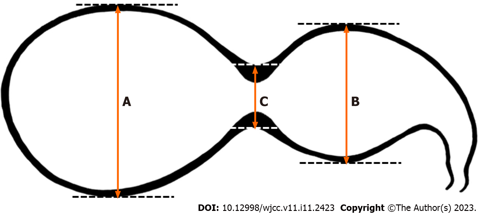
Figure 1 Schematic of the segmented gallbladder.
A: Long axis of the distal segment; B: Long axis of the proximal segment; C: Segmental luminal narrowing of the gallbladder (GB) body. Segmented GB was diagnosed if the sum of A and B was greater than 3 times C.
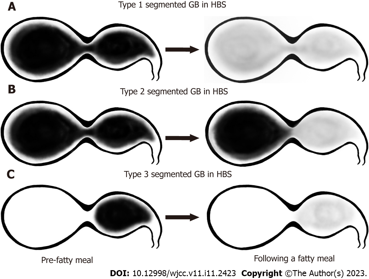
Figure 2 Schematic of the classification of filling and emptying patterns in segmented gallbladder as measured by hepatobiliary scintigraphy.
A: Type 1 was defined as a normal filling and emptying pattern; B: Type 2 was defined as an emptying defect at the distal segment; C: Type 3 was defined as a filling defect at the distal segment. GB: gallbladder; HBS: Hepatobiliary scintigraphy.
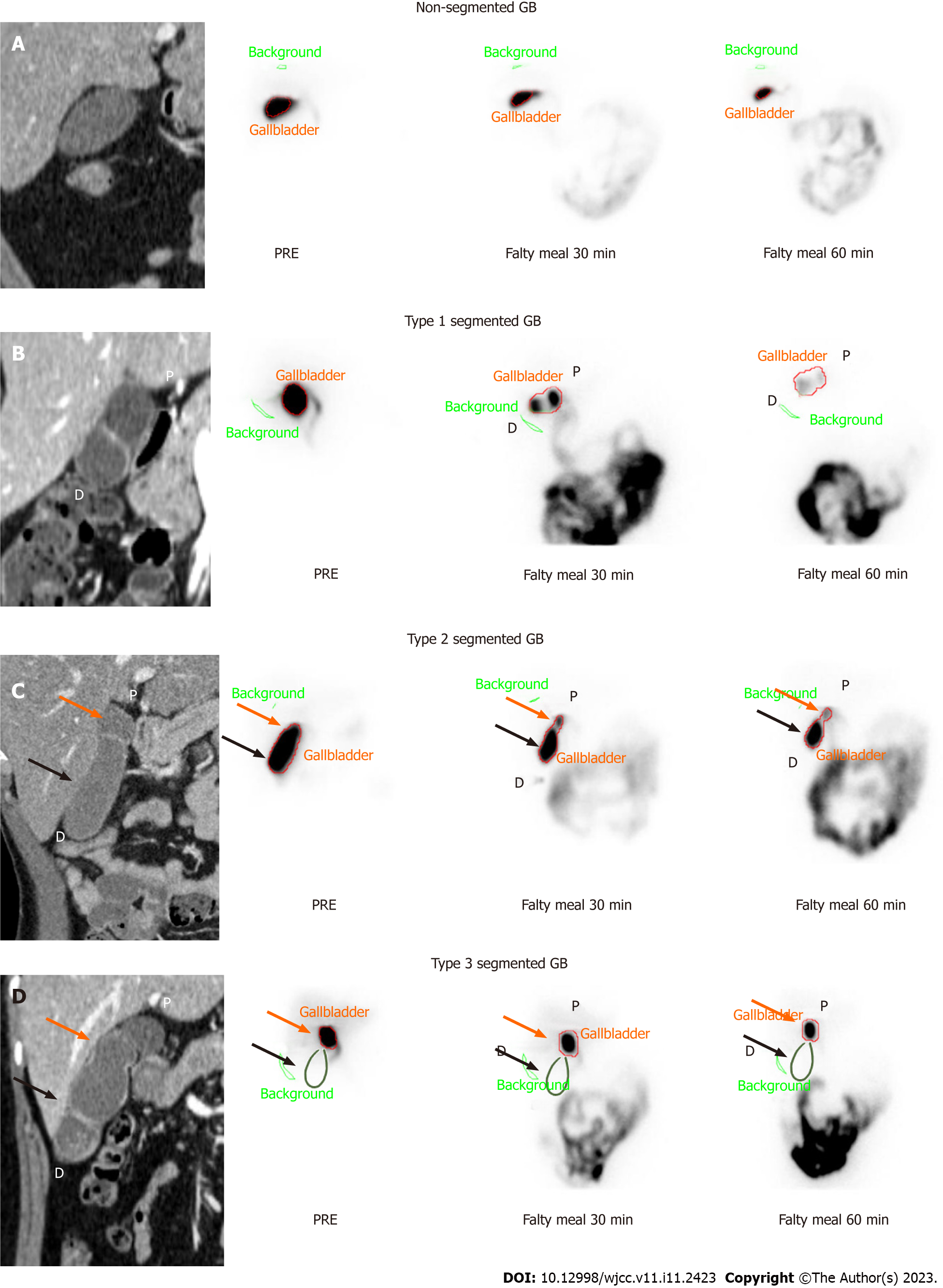
Figure 3 Typical images of computed tomography and hepatobiliary scintigraphy according to pattern in the normal and segmented gallbladder.
A: Hepatobiliary scintigraphy (HBS) in non-segmented gallbladder (GB). Reported GB ejection fraction (GBEF) was 74%; B: Type 1, normal filling and emptying pattern on HBS. Reported GBEF (95%) was reflected in both segments; C: Type 2, emptying defect of the distal segment on HBS. After 60 min, the proximal segment showed normal emptying, but the distal segment showed poor emptying. Reported GBEF (87%) reflects proximal segmentation more than distal segmentation; D: Type 3, filling defect of the distal segment on HBS. As the distal segment is not observed in the scanned image, other radiological validation is required. Reported GBEF (89%) reflects only proximal segmentation. The green line is a rough representation of the outline of the gallbladder per computed tomography. GB: gallbladder; P: Proximal segment (orange arrows); D: Distal segment (black arrows).
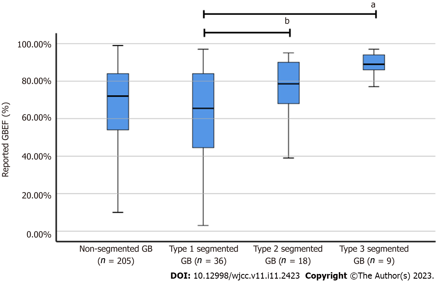
Figure 4 Gallbladder ejection fraction in non-segmented gallbladder and segmented gallbladder subtypes.
Reported gallbladder ejection fraction (GBEF) in non-segmented gallbladder (GB) was 67.40% ± 21.78%, whereas that of segmented GB was 62.50% ± 24.79% for Type 1, 75.89% ± 17.21% for Type 2, and 88.56% ± 7.20% for Type 3. aThere was a significant difference between reported GBEF of Types 1 and 3; bThe difference in GBEF of Types 1 and 2 was not statistically significant (P = 0.082). GB: Gallbladder; GBEF: Gallbladder ejection fraction.
- Citation: Lee YC, Jung WS, Lee CH, Kim SH, Lee SO. Classification of hepatobiliary scintigraphy patterns in segmented gallbladder according to anatomical discordance. World J Clin Cases 2023; 11(11): 2423-2434
- URL: https://www.wjgnet.com/2307-8960/full/v11/i11/2423.htm
- DOI: https://dx.doi.org/10.12998/wjcc.v11.i11.2423


