Copyright
©The Author(s) 2023.
World J Clin Cases. Apr 16, 2023; 11(11): 2423-2434
Published online Apr 16, 2023. doi: 10.12998/wjcc.v11.i11.2423
Published online Apr 16, 2023. doi: 10.12998/wjcc.v11.i11.2423
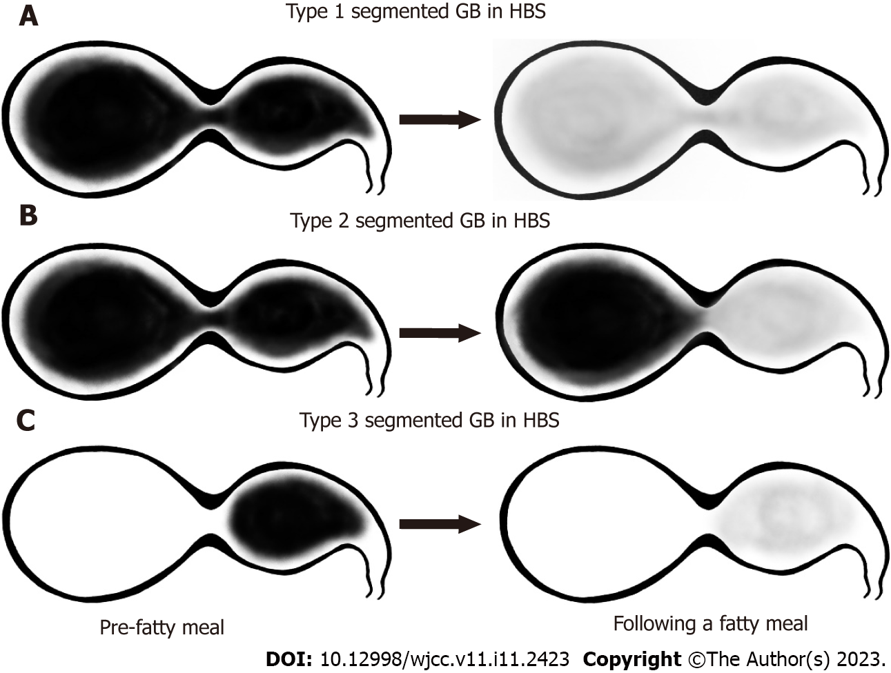
Figure 2 Schematic of the classification of filling and emptying patterns in segmented gallbladder as measured by hepatobiliary scintigraphy.
A: Type 1 was defined as a normal filling and emptying pattern; B: Type 2 was defined as an emptying defect at the distal segment; C: Type 3 was defined as a filling defect at the distal segment. GB: gallbladder; HBS: Hepatobiliary scintigraphy.
- Citation: Lee YC, Jung WS, Lee CH, Kim SH, Lee SO. Classification of hepatobiliary scintigraphy patterns in segmented gallbladder according to anatomical discordance. World J Clin Cases 2023; 11(11): 2423-2434
- URL: https://www.wjgnet.com/2307-8960/full/v11/i11/2423.htm
- DOI: https://dx.doi.org/10.12998/wjcc.v11.i11.2423


