Copyright
©The Author(s) 2022.
World J Clin Cases. Aug 6, 2022; 10(22): 7960-7967
Published online Aug 6, 2022. doi: 10.12998/wjcc.v10.i22.7960
Published online Aug 6, 2022. doi: 10.12998/wjcc.v10.i22.7960
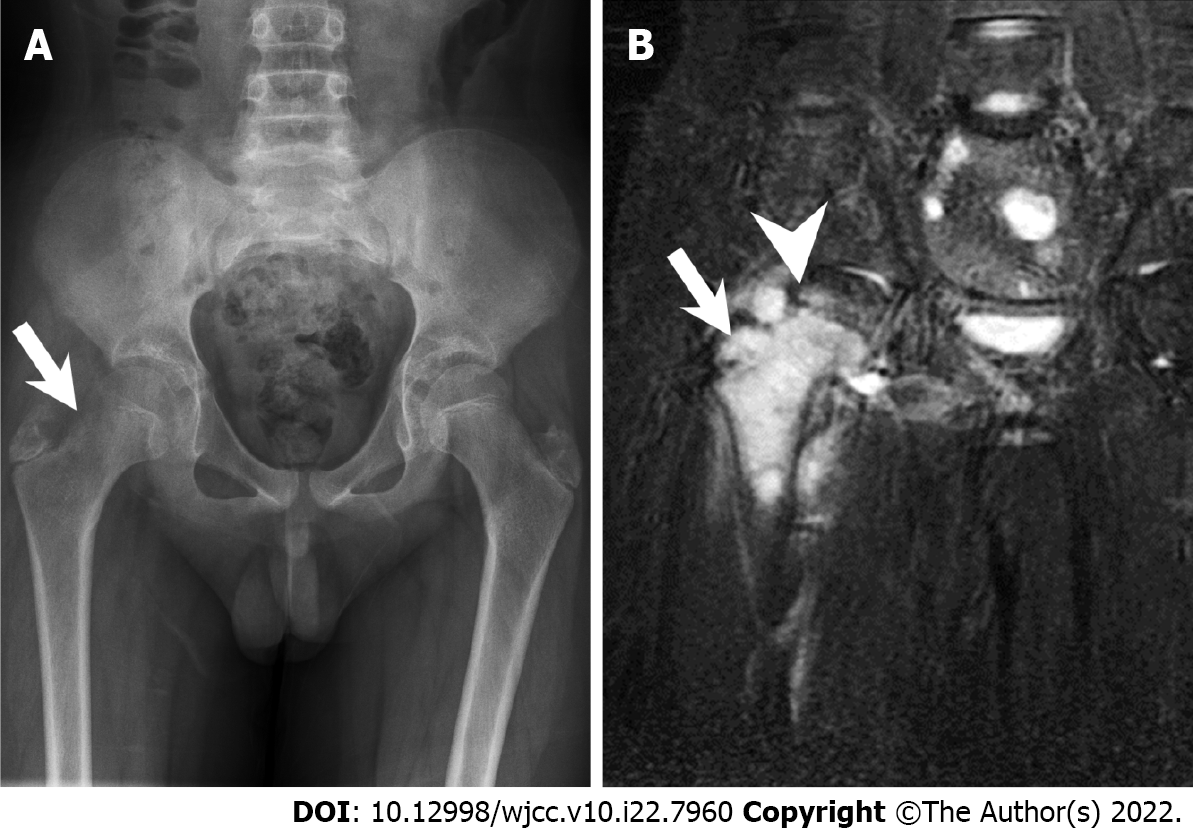
Figure 1 Radiography and magnetic resonance imaging.
A: Initial plain X-ray film of the pelvis (anteroposterior view). The arrow indicates the location of the tumor with cortical reaction at the femoral neck region; B: Initial pelvis coronary T2 STIR image showing involvement of the physis. The arrow and arrowhead indicate the location of the tumor and the femoral physis, respectively.
Figure 2 Initial chest computed tomography and chest computed tomography after neoadjuvant chemotherapy.
Full remission of the lung metastases after the neoadjuvant chemotherapy is noted. A: Initial: The metastatic Ewing’s sarcoma over the right middle lobe (arrowhead). Pulmonary artery is not distended (arrow), which implies no obstructive tumor thrombus in this multiple metastasis situation; B: After neoadjuvant chemotherapy: Localized same level chest computed tomography with the pulmonary artery (arrow). The previous tumor had disappeared; C: Initial: The metastatic Ewing’s sarcoma in the right inferior lobe (arrowhead) and left inferior lobe with another metastatic Ewing’s sarcoma (arrowhead); D: After neoadjuvant chemotherapy: Localized same level chest computed tomography with similar pulmonary artery distribution. The previous tumors had disappeared.
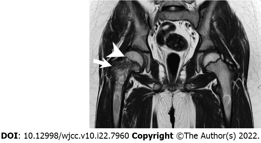
Figure 3 T2 magnetic resonance image (coronary view) after neoadjuvant chemotherapy.
The arrow and arrowhead indicate the location of the tumor and the femoral physis, respectively. Note that the tumor is now confined only to the physis.
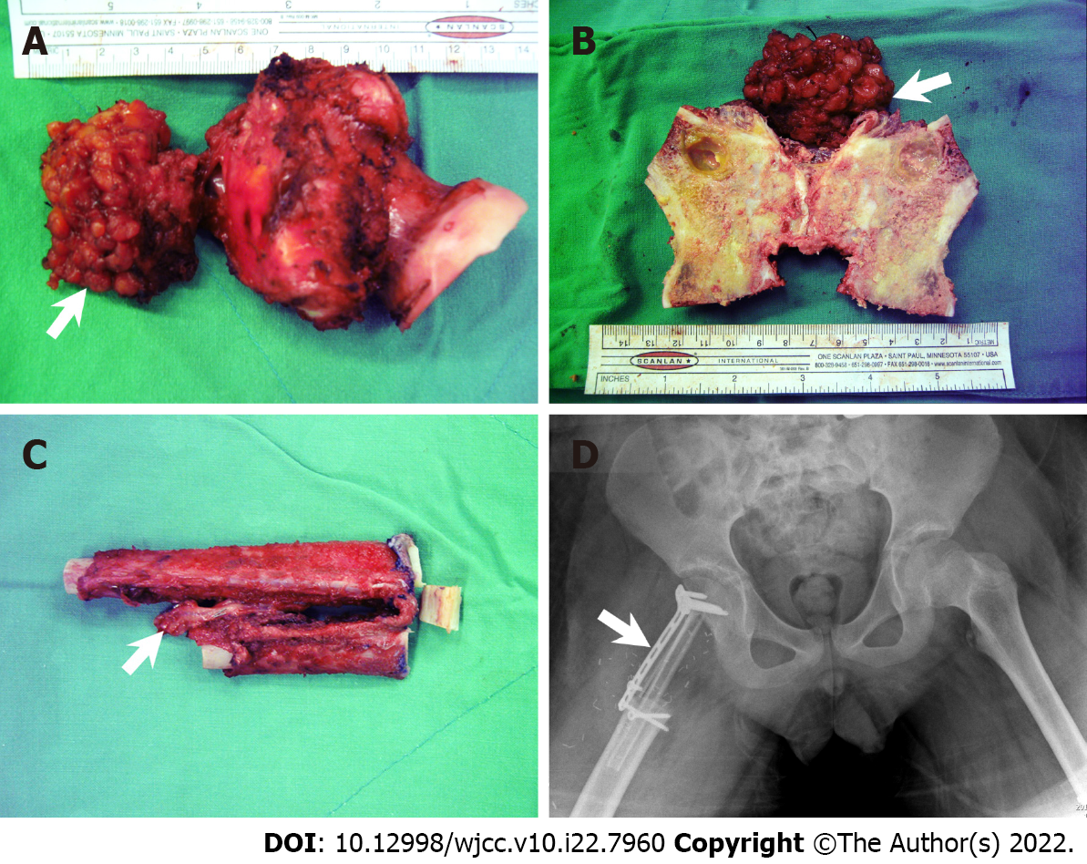
Figure 4 Intraoperative sample and postoperative pelvic plain imaging.
A: Gross view of the resected tumor and resected femoral neck (arrow: Ewing’s sarcoma extra-bony part); B: Split resected femoral neck; C: Folded autologous fibular graft (arrow: vascular pedicle); D: Pelvic plain imaging (anteroposterior) obtained immediately after the surgery (arrow: locking plate used to fix the fibular graft).
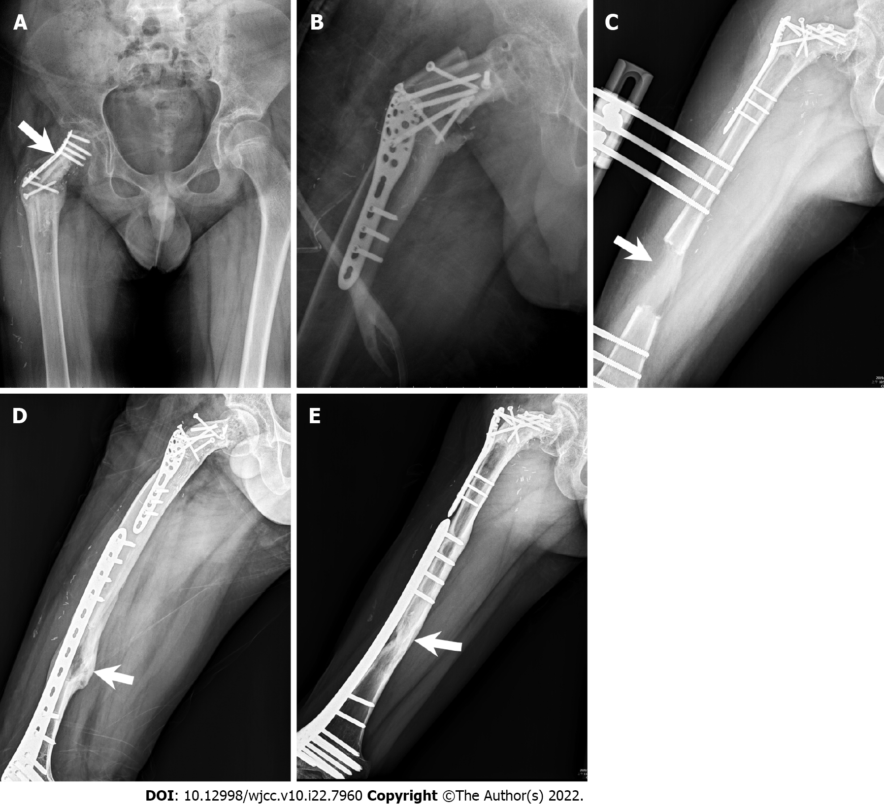
Figure 5 Radiographic images.
A: Plain X-ray film obtained after an increase in partial weight bearing. The plate was deformed (arrow: bending site of the plate); B: Revision and fixation with Arbeitsgemeinschaft für Osteosynthesefragen proximal humeral internal locking system plate; C: Radiographic image of orthosis with complete distraction. The femoral distraction gap is approximately 6.5 cm (arrow: distraction gap); D: Radiographic image showing removal of the orthosis and international fixation with less-invasive stabilization system complete corticalization (arrow: corticalized bone); E: Radiographic image showing complete consolidation phase (arrow: cordialized bone with increased radiopacity).
- Citation: Lai CY, Chen KJ, Ho TY, Li LY, Kuo CC, Chen HT, Fong YC. Resection with limb salvage in an Asian male adolescent with Ewing’s sarcoma: A case report. World J Clin Cases 2022; 10(22): 7960-7967
- URL: https://www.wjgnet.com/2307-8960/full/v10/i22/7960.htm
- DOI: https://dx.doi.org/10.12998/wjcc.v10.i22.7960


