Copyright
©The Author(s) 2022.
World J Clin Cases. Aug 6, 2022; 10(22): 7960-7967
Published online Aug 6, 2022. doi: 10.12998/wjcc.v10.i22.7960
Published online Aug 6, 2022. doi: 10.12998/wjcc.v10.i22.7960
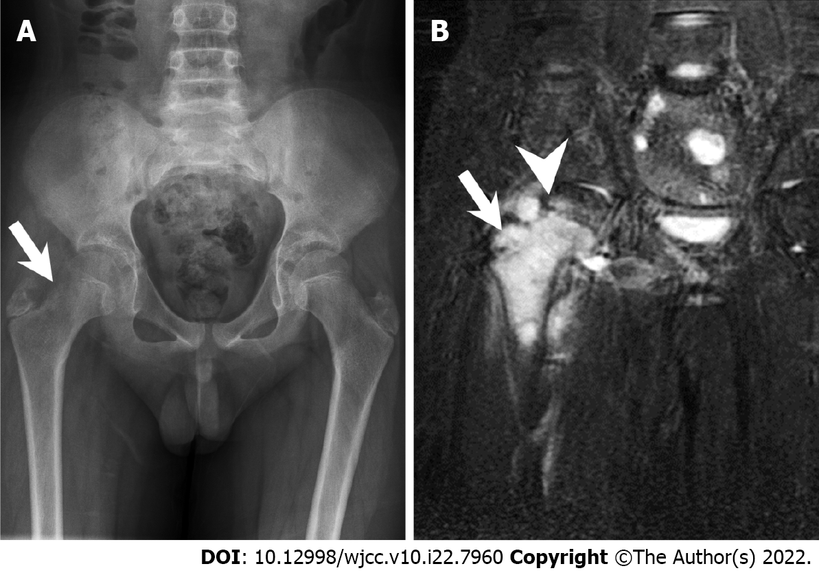
Figure 1 Radiography and magnetic resonance imaging.
A: Initial plain X-ray film of the pelvis (anteroposterior view). The arrow indicates the location of the tumor with cortical reaction at the femoral neck region; B: Initial pelvis coronary T2 STIR image showing involvement of the physis. The arrow and arrowhead indicate the location of the tumor and the femoral physis, respectively.
- Citation: Lai CY, Chen KJ, Ho TY, Li LY, Kuo CC, Chen HT, Fong YC. Resection with limb salvage in an Asian male adolescent with Ewing’s sarcoma: A case report. World J Clin Cases 2022; 10(22): 7960-7967
- URL: https://www.wjgnet.com/2307-8960/full/v10/i22/7960.htm
- DOI: https://dx.doi.org/10.12998/wjcc.v10.i22.7960


