Copyright
©The Author(s) 2022.
World J Clin Cases. Jun 26, 2022; 10(18): 6254-6260
Published online Jun 26, 2022. doi: 10.12998/wjcc.v10.i18.6254
Published online Jun 26, 2022. doi: 10.12998/wjcc.v10.i18.6254
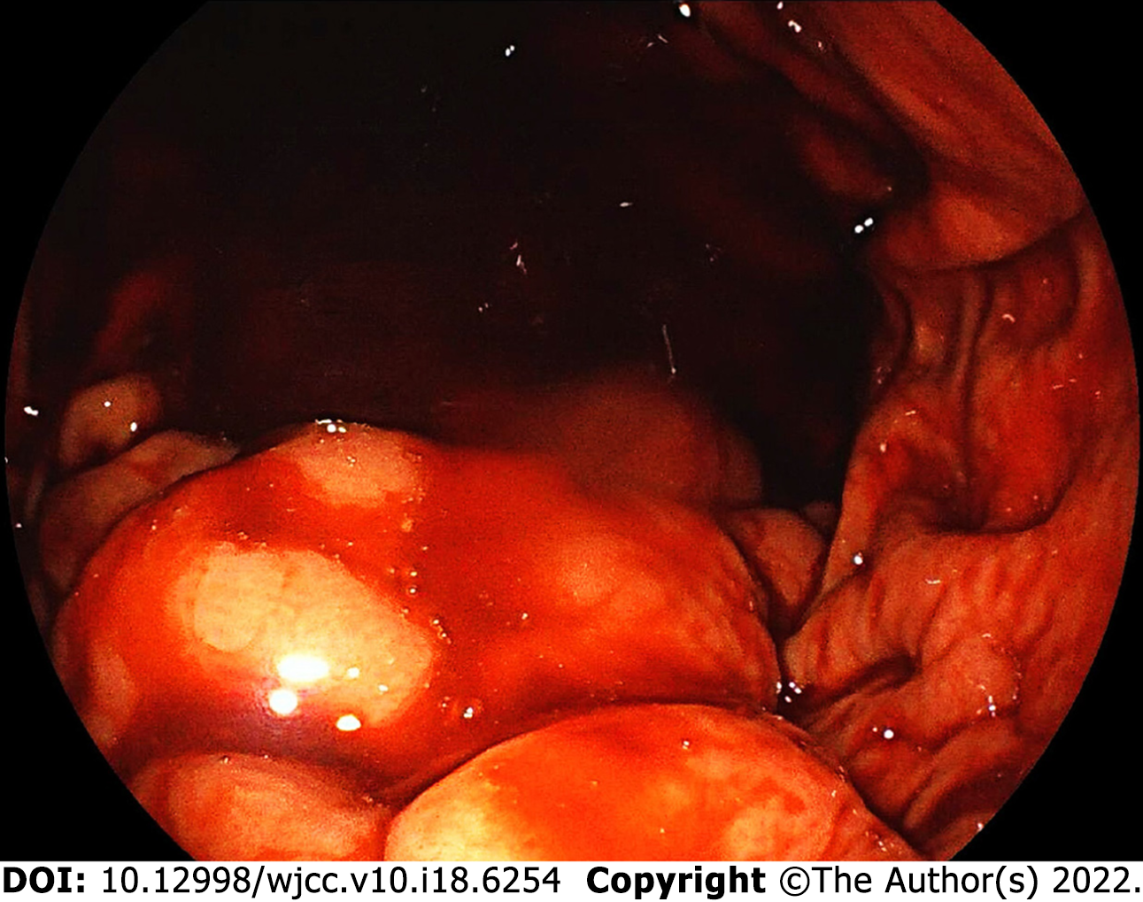
Figure 1 Gastroscopic image.
Gastroscopy revealed gastric variceal with signs of recent bleeding in the absence of active bleeding.
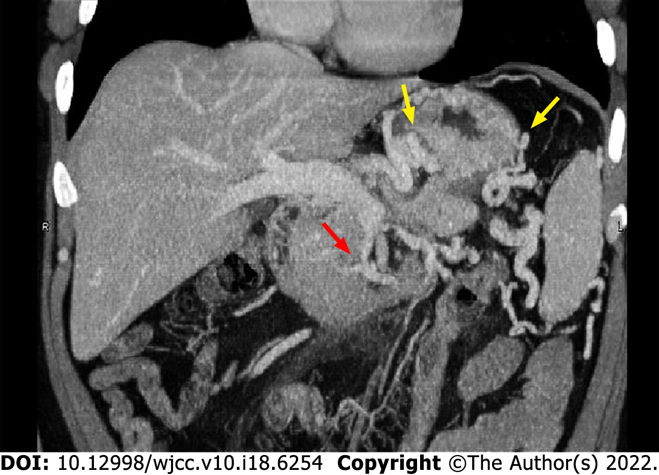
Figure 2 Abdominal computed tomography venography image.
Computed tomography venography revealed stenosis of the proximal superior mesenteric vein (red arrow), invisible proximal splenic vein, and increased collateral circulations (yellow arrows).
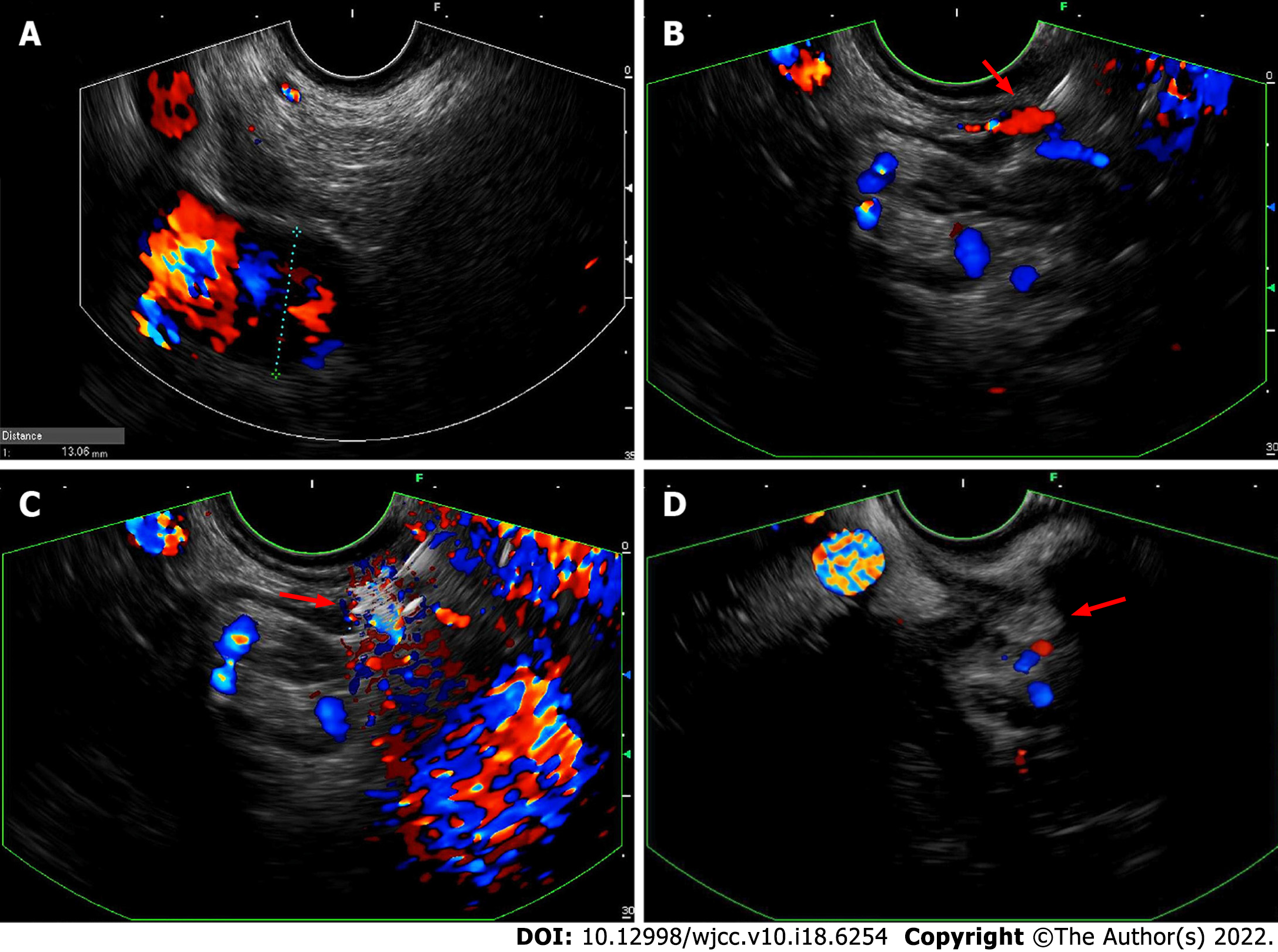
Figure 3 Endoscopic ultrasound images.
A: Endoscopic ultrasound revealed an enlarged portal vein; B: A confluence of gastric varices was identified and selected as the injection site (red arrow); C: Undiluted N-butyl-2-cyanoacrylate (red arrow) was injected into the selected gastric varix via a 22-gauge needle; D: Hyperechoic fillings (red arrow) and decreased blood flow signals were observed after injections.
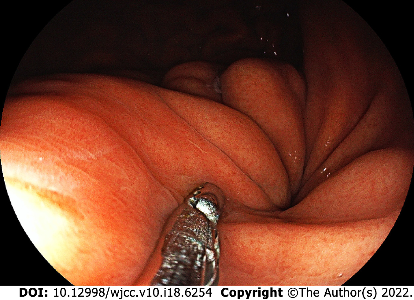
Figure 4 Gastroscopic image.
With the help of biopsy forceps, the follow-up gastroscopy revealed firm gastric submucosa and no sign of N-butyl-2-cyanoacrylate expulsion.
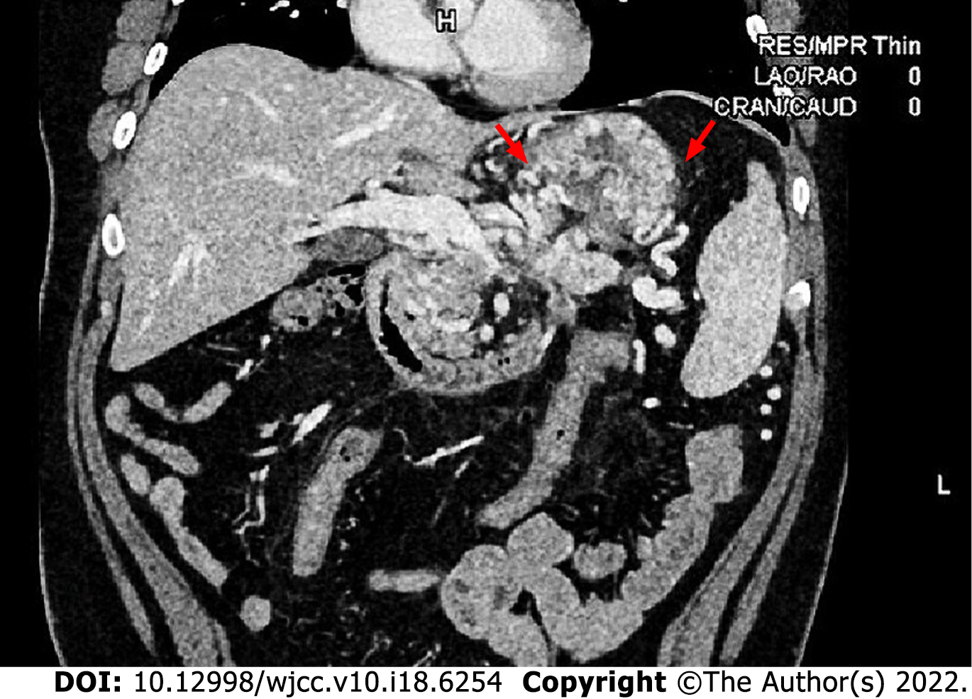
Figure 5 Computed tomography venography image.
Compared with the results before the operation (Figure 2), follow-up computed tomography venography revealed improvements in left-sided portal hypertension and collateral circulations (red arrows).
- Citation: Yang J, Zeng Y, Zhang JW. Modified endoscopic ultrasound-guided selective N-butyl-2-cyanoacrylate injections for gastric variceal hemorrhage in left-sided portal hypertension: A case report. World J Clin Cases 2022; 10(18): 6254-6260
- URL: https://www.wjgnet.com/2307-8960/full/v10/i18/6254.htm
- DOI: https://dx.doi.org/10.12998/wjcc.v10.i18.6254


