Copyright
©The Author(s) 2022.
World J Clin Cases. Jun 16, 2022; 10(17): 5869-5876
Published online Jun 16, 2022. doi: 10.12998/wjcc.v10.i17.5869
Published online Jun 16, 2022. doi: 10.12998/wjcc.v10.i17.5869
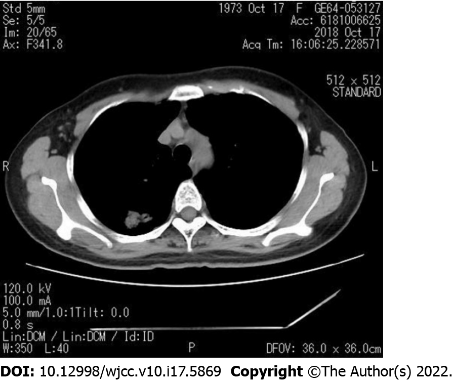
Figure 1 Computerized tomography examination showing a mass located in the posterior segment of the right upper lobe of the lung.
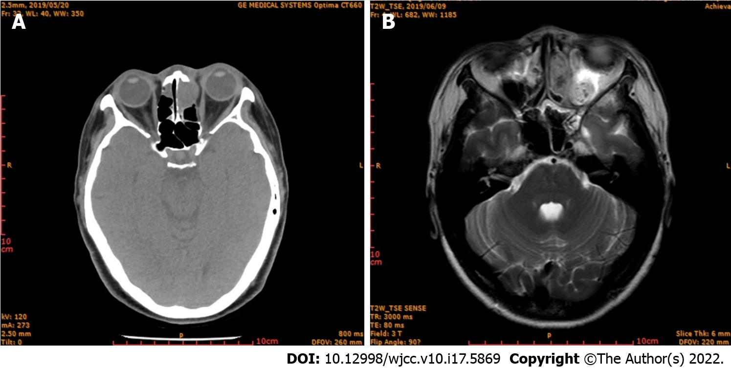
Figure 2 Imaging examination.
A: Computerized tomography examination showing tumor invasion of the ethmoid sinus; B: Magnetic resonance imaging showing a mass in the ethmoid sinus.
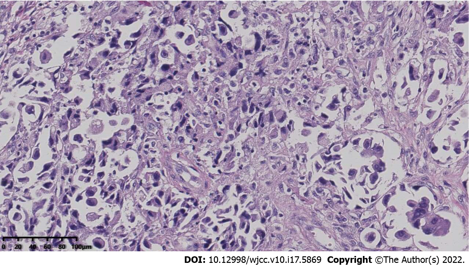
Figure 3 Pathological immunohistochemistry showing adenocarcinoma infiltrates between fibrous connective tissues, indicating lung adenocarcinoma.
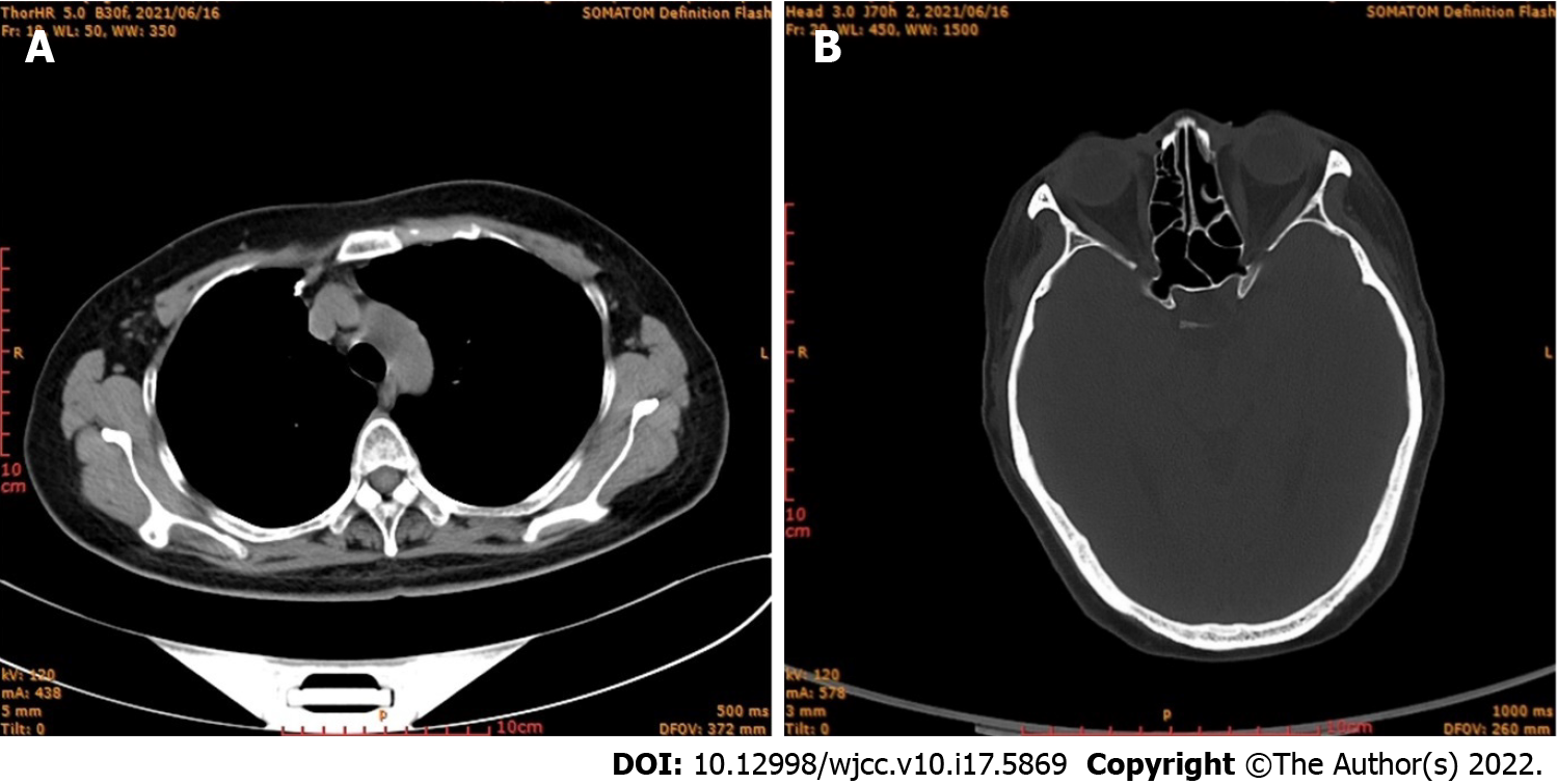
Figure 4 Computed tomography images.
A: Computed tomography (CT) of lung obtained two years later, showing no recurrence; B: CT of paranasal sinus obtained two years later, showing no recurrence.
- Citation: Li WJ, Xue HX, You JQ, Chao CJ. Lung adenocarcinoma metastasis to paranasal sinus: A case report . World J Clin Cases 2022; 10(17): 5869-5876
- URL: https://www.wjgnet.com/2307-8960/full/v10/i17/5869.htm
- DOI: https://dx.doi.org/10.12998/wjcc.v10.i17.5869


