Copyright
©The Author(s) 2022.
World J Clin Cases. May 6, 2022; 10(13): 4288-4293
Published online May 6, 2022. doi: 10.12998/wjcc.v10.i13.4288
Published online May 6, 2022. doi: 10.12998/wjcc.v10.i13.4288
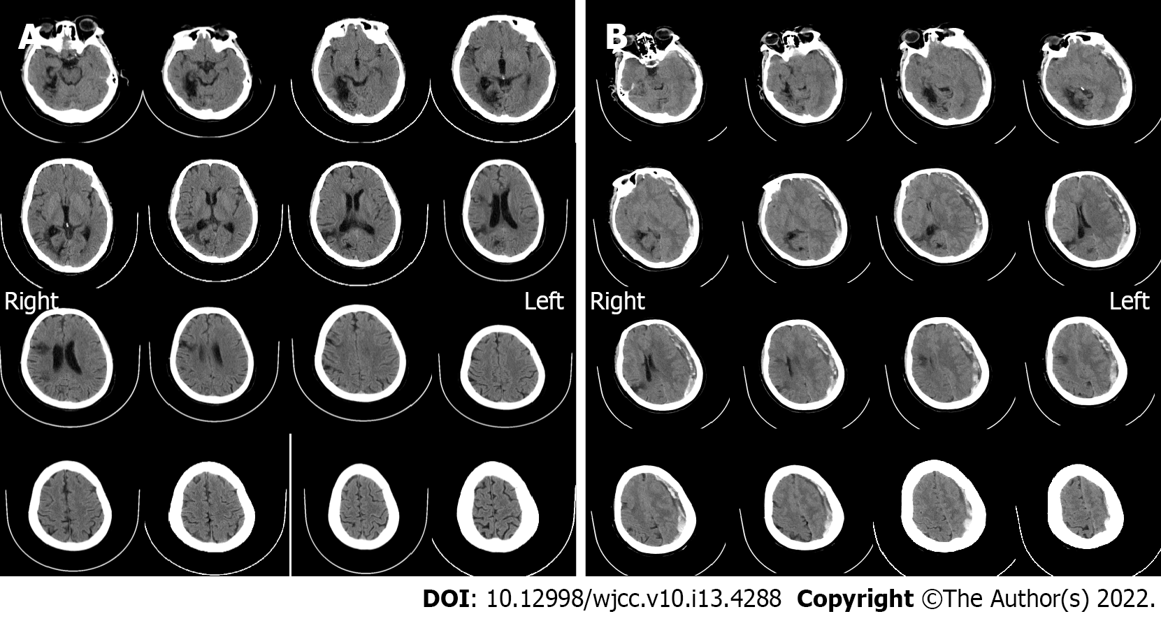
Figure 1 Imaging examinations.
A: Computed tomography (CT) scan on admission (October 10, 2021). It showed no hematoma, but just degenerative changes indicative of past cerebral infarction; B: Emergency CT findings before the craniotomy (October 19, 2021). Acute subdural hematoma with a prominent mass effect was found, with some low-density components inside it. The midline was shifted indicating brain hernia.
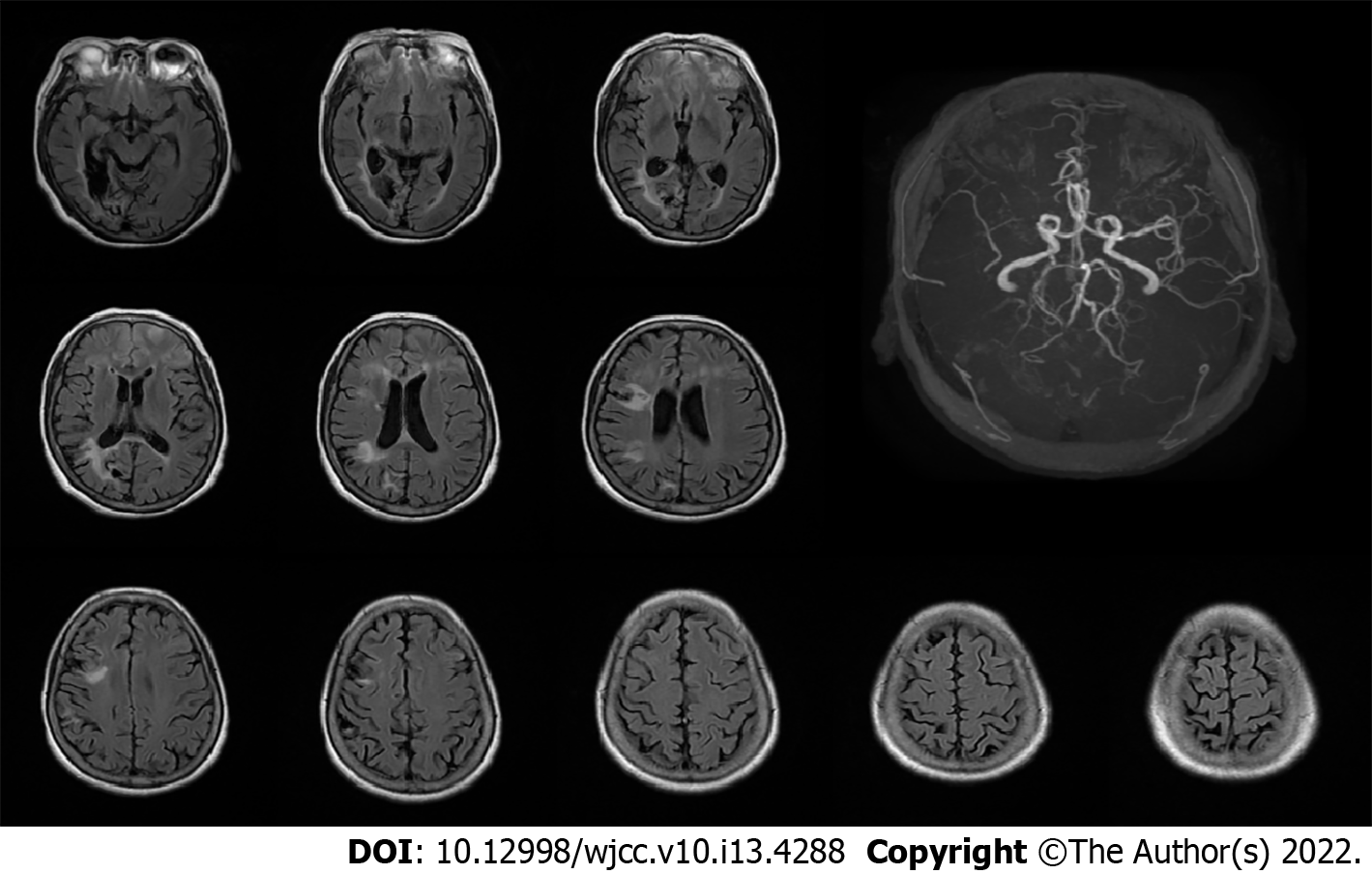
Figure 2 Magnetic resonance imaging on day 3 of treatment (October 12, 2021).
The scan indicated no hemorrhage events, while an magnetic resonance angiography showed no development of right middle cerebral artery (white arrow).
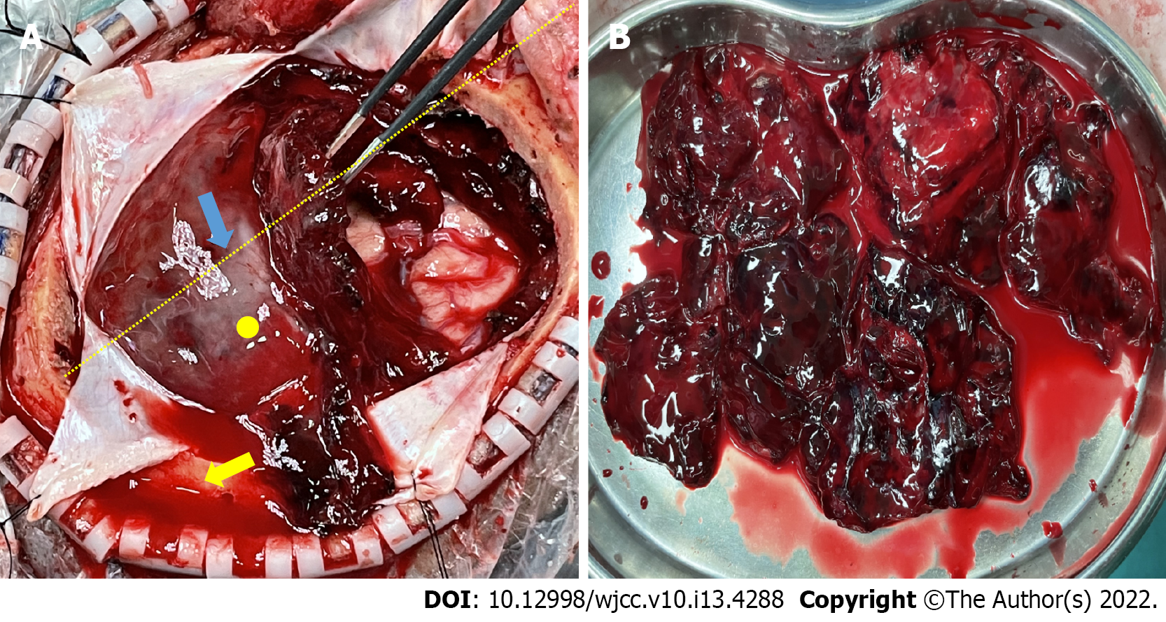
Figure 3 Intraoperative findings.
A: Initial attempt to evacuate the subdural hematoma. The yellow dotted line indicates the lateral fissure, thickened membranes were found over the hematoma’s surface (blue arrow), the yellow circle is the point where we found a minor arterial bleeding after the mass was removed. The yellow arrow shows epidural bloody fluids that were due to the absence of dural suspension because we wanted to decompress it as quickly as possible and later suspend the dura. The hematoma itself contained hardly any liquefied contents; B: Completed removal of the hematoma in pieces. The inner membrane was not as thick as the outer one.
- Citation: Lv HT, Zhang LY, Wang XT. Enigmatic rapid organization of subdural hematoma in a patient with epilepsy: A case report. World J Clin Cases 2022; 10(13): 4288-4293
- URL: https://www.wjgnet.com/2307-8960/full/v10/i13/4288.htm
- DOI: https://dx.doi.org/10.12998/wjcc.v10.i13.4288


