Copyright
©The Author(s) 2022.
World J Clin Cases. May 6, 2022; 10(13): 4288-4293
Published online May 6, 2022. doi: 10.12998/wjcc.v10.i13.4288
Published online May 6, 2022. doi: 10.12998/wjcc.v10.i13.4288
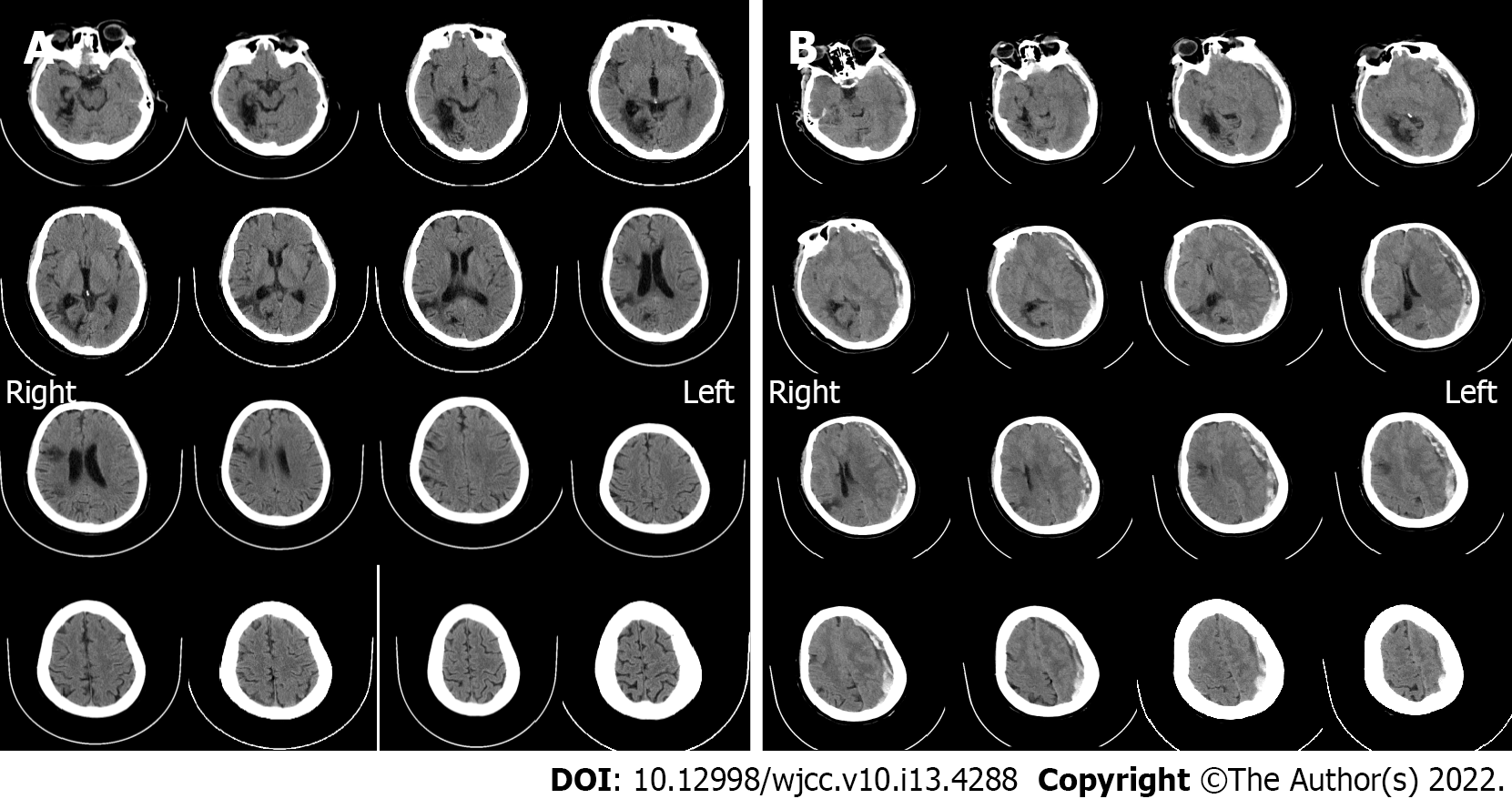
Figure 1 Imaging examinations.
A: Computed tomography (CT) scan on admission (October 10, 2021). It showed no hematoma, but just degenerative changes indicative of past cerebral infarction; B: Emergency CT findings before the craniotomy (October 19, 2021). Acute subdural hematoma with a prominent mass effect was found, with some low-density components inside it. The midline was shifted indicating brain hernia.
- Citation: Lv HT, Zhang LY, Wang XT. Enigmatic rapid organization of subdural hematoma in a patient with epilepsy: A case report. World J Clin Cases 2022; 10(13): 4288-4293
- URL: https://www.wjgnet.com/2307-8960/full/v10/i13/4288.htm
- DOI: https://dx.doi.org/10.12998/wjcc.v10.i13.4288


