Copyright
©The Author(s) 2017.
World J Methodol. Dec 26, 2017; 7(4): 139-147
Published online Dec 26, 2017. doi: 10.5662/wjm.v7.i4.139
Published online Dec 26, 2017. doi: 10.5662/wjm.v7.i4.139
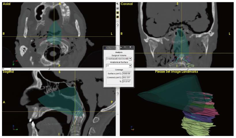
Figure 7 Detail of ApproachViewer screen during post-dissection analyses.
An endonasal endoscopic approach to the odontoid was performed in the anatomy laboratory and is retrieved and visualized in the three axes and as a volume. Different areas of interest have been contoured at the level of the anterior craniovertebral junction. The box at the center shows the absolute and percentage value of contoured C1 exposed by the approach (see online additional material - ApproachViewer guide 1.0 for further details).
- Citation: Doglietto F, Qiu J, Ravichandiran M, Radovanovic I, Belotti F, Agur A, Zadeh G, Fontanella MM, Kucharczyk W, Gentili F. Quantitative comparison of cranial approaches in the anatomy laboratory: A neuronavigation based research method. World J Methodol 2017; 7(4): 139-147
- URL: https://www.wjgnet.com/2222-0682/full/v7/i4/139.htm
- DOI: https://dx.doi.org/10.5662/wjm.v7.i4.139


