Copyright
©The Author(s) 2016.
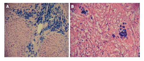
Figure 5 Non homogeneous iron distribution in the liver and spleen of an iron loaded thalassaemia patient.
Liver and spleen biopsy photographs (× 20) of a 29-year-old, 55 kg male thalassaemia patient. The liver biopsy was obtained during splenectomy. A: Liver section showing non unifom iron deposition stained with Pearl’s Prussian blue. There are hemosiderin deposits in hepatocytes and Kupffer cells and especially within bile ducts; B: Spleen section where iron deposits were stained with Pearl’s Prussian blue. There are non uniform hemosiderin deposits within cytoplasma and nucleus of macrophages. Four months before the splenectomy the patient had an MRI T2* (ms) of heart 4.1, liver 0.0, spleen 2.9, and serum ferritin of 3850 μg/L. Adapted from ref. [96]. MRI: Magnetic resonance imaging.
- Citation: Kontoghiorghe CN, Kontoghiorghes GJ. New developments and controversies in iron metabolism and iron chelation therapy. World J Methodol 2016; 6(1): 1-19
- URL: https://www.wjgnet.com/2222-0682/full/v6/i1/1.htm
- DOI: https://dx.doi.org/10.5662/wjm.v6.i1.1


