
















Copyright: ©Author(s) 2026.
World J Methodol. Sep 20, 2026; 16(3): 120949
Published online Sep 20, 2026. doi: 10.5662/wjm.120949
Published online Sep 20, 2026. doi: 10.5662/wjm.120949
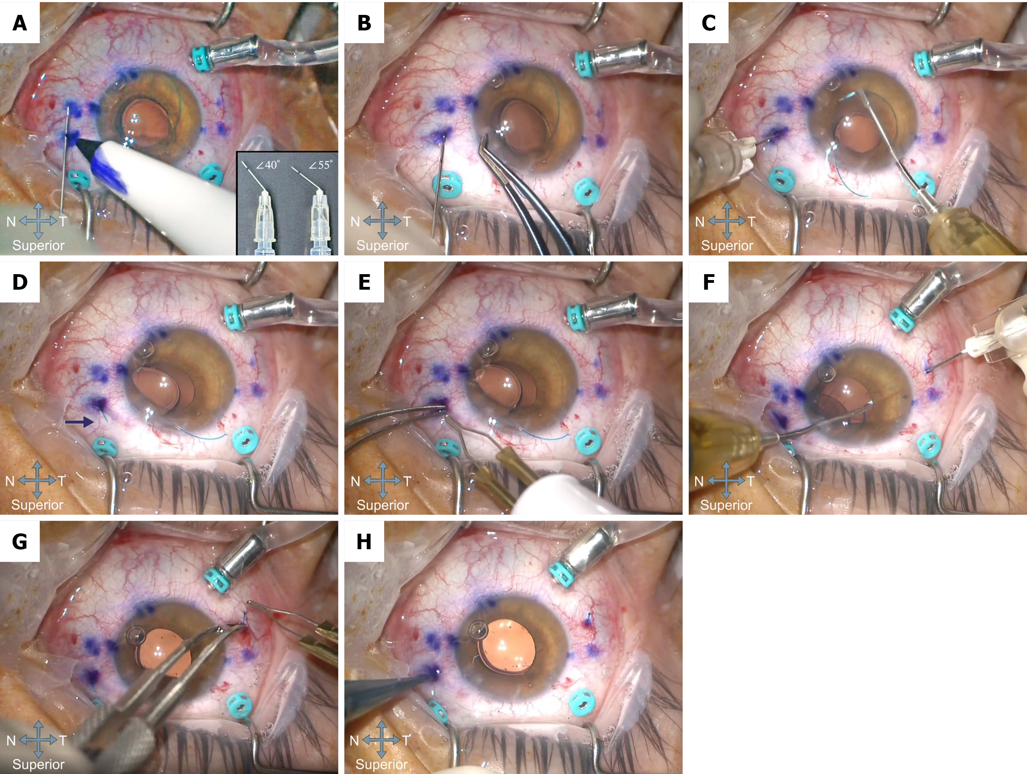
Figure 2 Stepwise illustration of the Yamane technique for flanged intrascleral haptic fixation of a posterior chamber intraocular lens in a right eye, performed in conjunction with pars plana vitrectomy.
Trocar cannulas (teal) for 25-gauge pars plana vitrectomy ports placed prior to intraocular lens (IOL) fixation to allow complete vitreous clearance and to reduce the risk of postoperative traction and cystoid macular edema are visible throughout panels A-H; A: Needle marking and needle flexion planning. Blue marking ink dots are visible on the conjunctiva, identifying the planned 30-gauge thin-wall needle entry sites (2.0 mm posterior to the limbus); a reference mark is placed on the needle shaft to guide the intended intrascleral tunnel length (approximately 2.0-2.5 mm); pre-bent 30-gauge thin-wall needles are prepared with different flexion angles (inset: Needle #1: Approximately 40°; needle #2: Approximately 55°); B: Transscleral needle entry; C: Haptic docking into the needle lumen; D: Externalization of the haptic through the scleral tunnel, the externalized haptic is highlighted by an arrow at the scleral exit site; E: Flange formation of the first haptic by low-temperature cautery; F: Externalization of the second haptic and symmetric positioning; G: Flange formation of the second haptic by low-temperature cautery; H: Burial of the flanged haptics and final IOL centration. T: Temporal; N: Nasal.
- Citation: Usta Küçükbezirci G, Gürcan B, Patel N, Ramsey DJ, Alwreikat A. Standardizing the Yamane flanged intrascleral haptic fixation: A framework for efficacy, complication mitigation, and reproducible training. World J Methodol 2026; 16(3): 120949
- URL: https://www.wjgnet.com/2222-0682/full/v16/i3/120949.htm
- DOI: https://dx.doi.org/10.5662/wjm.120949