
















Copyright: ©Author(s) 2026.
World J Methodol. Sep 20, 2026; 16(3): 120949
Published online Sep 20, 2026. doi: 10.5662/wjm.120949
Published online Sep 20, 2026. doi: 10.5662/wjm.120949
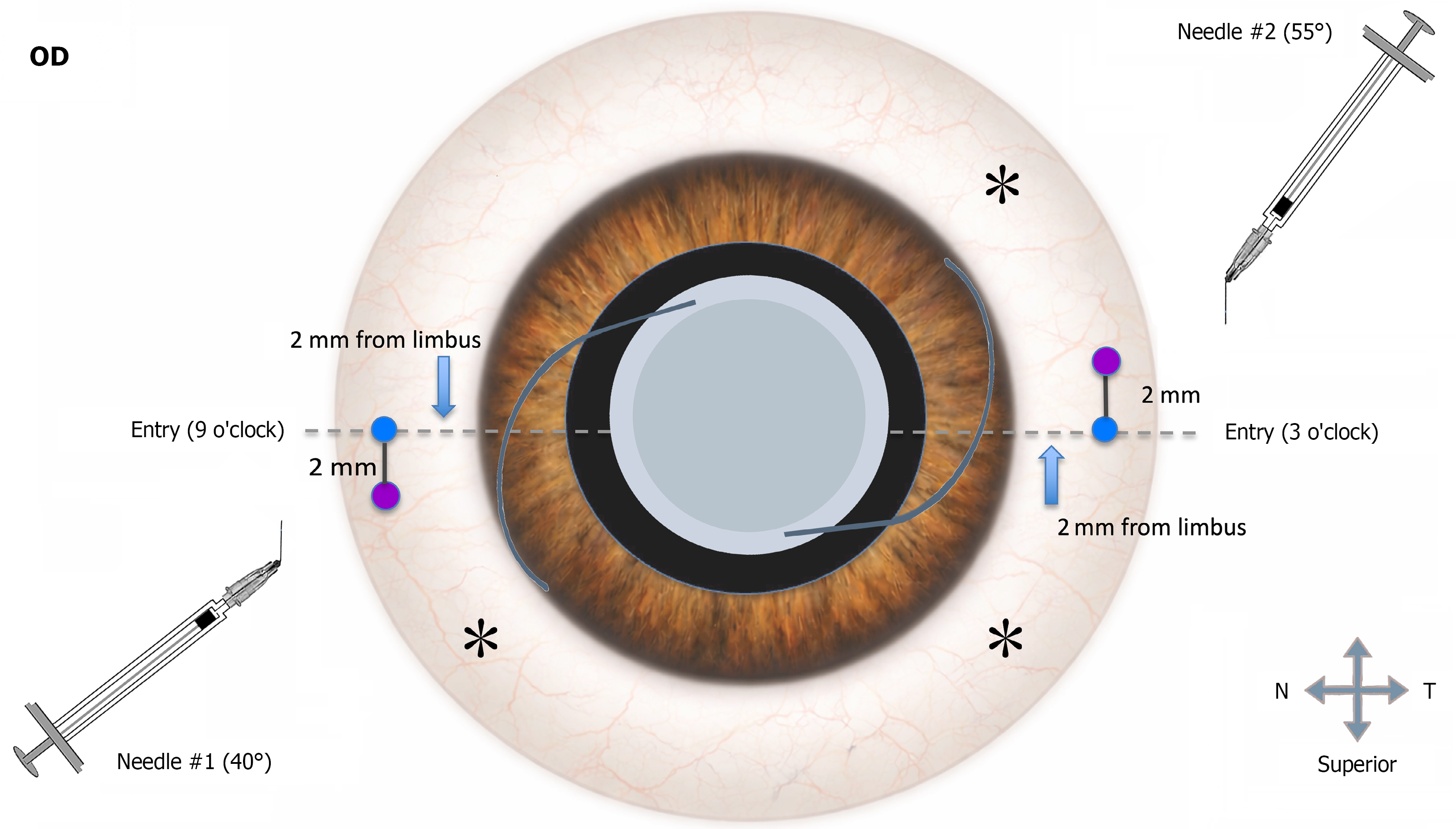
Figure 1 Diagram of Yamane flanged intrascleral intraocular lens fixation technique in a right eye.
A sterile surgical marking pen is used intraoperatively to identify the scleral fixation sites at the 3 and 9 o’clock meridians. The scleral entry points (purple dots) are marked at 2.0 mm (2 mm to 2.5 mm) posterior to the limbus to guide symmetric tunnel creation. Pre-bent 30-gauge thin-wall needles with different flexion angles (needle #1: Approximately 40°; needle #2: Approximately 55°) are prepared to facilitate ergonomic handling and controlled transscleral entry. The needles are then advanced toward the planned intraocular penetration points (blue dots) along an intrascleral trajectory (approximately 20-30°), creating tunnels of about 2.0-2.5 mm before entering the posterior chamber. This configuration facilitates controlled haptic externalization and stable posterior chamber intraocular lens fixation. Asterisks (*) indicate the three pars plana vitrectomy port entry sites: One superotemporal infusion cannula and two instrument ports placed approximately 3.5 mm posterior to the limbus in the inferotemporal and superior quadrants, respectively. T: Temporal; N: Nasal; OD: Oculus dexter (right eye).
- Citation: Usta Küçükbezirci G, Gürcan B, Patel N, Ramsey DJ, Alwreikat A. Standardizing the Yamane flanged intrascleral haptic fixation: A framework for efficacy, complication mitigation, and reproducible training. World J Methodol 2026; 16(3): 120949
- URL: https://www.wjgnet.com/2222-0682/full/v16/i3/120949.htm
- DOI: https://dx.doi.org/10.5662/wjm.120949