
















©The Author(s) 2026.
World J Methodol. Mar 20, 2026; 16(1): 112458
Published online Mar 20, 2026. doi: 10.5662/wjm.v16.i1.112458
Published online Mar 20, 2026. doi: 10.5662/wjm.v16.i1.112458
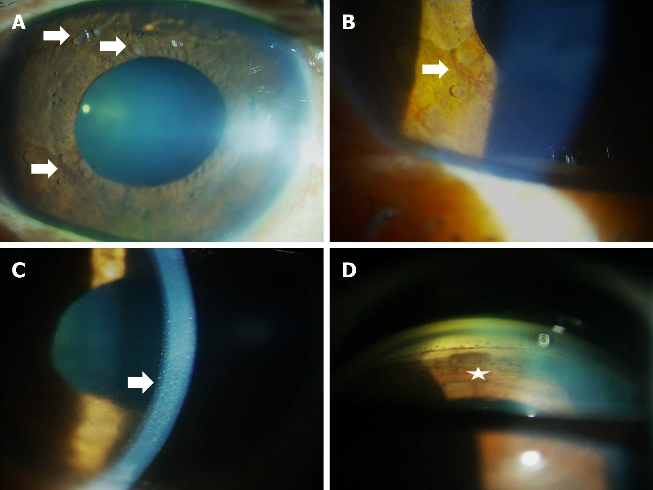
Figure 2 Iris nodules at presentation (Case 2).
A: Multiple semi-transparent, jelly like nodules were found in multiple quadrants (arrows); B: Neova
- Citation: Nayak B, Mohapatra PR, Chakraborty K, Nanda J, Haripriya S, Mantha SP, Sethy M, Panda BB. Are iris masses in lung carcinoma always a metastasis: Two case reports. World J Methodol 2026; 16(1): 112458
- URL: https://www.wjgnet.com/2222-0682/full/v16/i1/112458.htm
- DOI: https://dx.doi.org/10.5662/wjm.v16.i1.112458