
















©The Author(s) 2026.
World J Methodol. Mar 20, 2026; 16(1): 112458
Published online Mar 20, 2026. doi: 10.5662/wjm.v16.i1.112458
Published online Mar 20, 2026. doi: 10.5662/wjm.v16.i1.112458
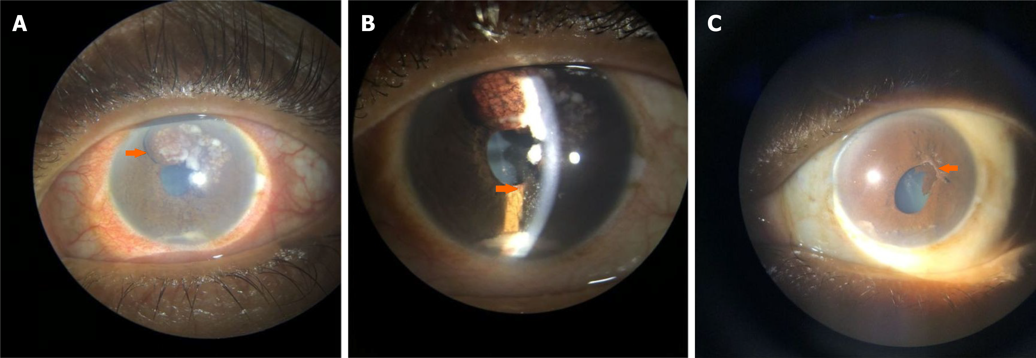
Figure 1 Case 1.
A: Slit-lamp bio-microscopy image of the left eye showing congestion and iris mass from 11 o'clock to 1 o'clock (orange arrow) with whitish flocculated materials deposition over iris and in the anterior chamber; B: Higher magnification (16 ×) image showing dispersed whitish flocculent material in the anterior chamber and neovascularization of iris (arrow); C: Post chemotherapy appearance of the left eye shows the disappearance of the iris mass and the flocculent materials, with post-inflammatory ectropion uvea and posterior synechiae from 12 o'clock to 3 o'clock (orange arrow).
- Citation: Nayak B, Mohapatra PR, Chakraborty K, Nanda J, Haripriya S, Mantha SP, Sethy M, Panda BB. Are iris masses in lung carcinoma always a metastasis: Two case reports. World J Methodol 2026; 16(1): 112458
- URL: https://www.wjgnet.com/2222-0682/full/v16/i1/112458.htm
- DOI: https://dx.doi.org/10.5662/wjm.v16.i1.112458