
















©The Author(s) 2026.
World J Methodol. Mar 20, 2026; 16(1): 107426
Published online Mar 20, 2026. doi: 10.5662/wjm.v16.i1.107426
Published online Mar 20, 2026. doi: 10.5662/wjm.v16.i1.107426
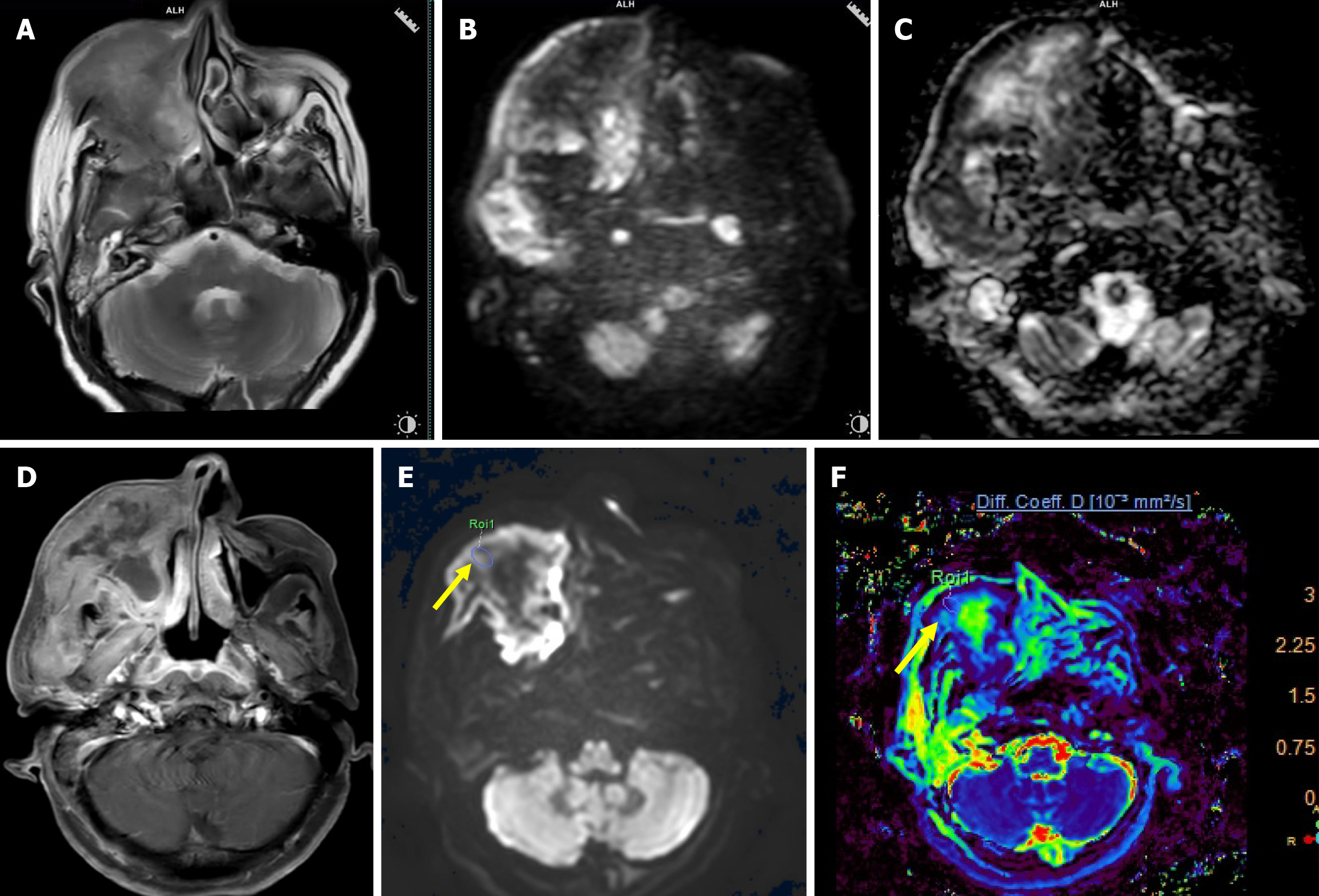
Figure 3 A 55-year-old female with undifferentiated carcinoma of the right maxillary sinus.
A: Axial T2-weighted image (T2WI) shows an ill-defined intermediate signal intensity mass epicentered in the right maxillary sinus with infiltration of the subcutaneous plane and skin; B: High b-value (b = 1000 seconds/mm2) image shows areas of high signal intensity; C: On corresponding apparent diffusion coefficient (ADC) map, similar areas showed hypointensity suggestive of true diffusion restriction (mean ADC value of 0.77 × 10⁻³ mm2/second); D: Axial post contrast T1WI image shows heterogeneous enhancement with central nonenhancing areas; E: High b-value image (b = 2000 seconds/mm2) of intravoxel incoherent motion imaging/diffusion kurtosis imaging sequence with region of interest (ROI) drawn (arrow) over the mass; F: True diffusion coefficient map after drawing ROI showed low value (0.68 × 10⁻³ mm2/second).
- Citation: Saini M, Manchanda S, Bhalla AS, Kandasamy D, Kakkar A, Thakar A. Multiparametric magnetic resonance imaging in differentiating benign and malignant sinonasal masses: A prospective study and literature review. World J Methodol 2026; 16(1): 107426
- URL: https://www.wjgnet.com/2222-0682/full/v16/i1/107426.htm
- DOI: https://dx.doi.org/10.5662/wjm.v16.i1.107426