Copyright
©The Author(s) 2016.
World J Transl Med. Apr 12, 2016; 5(1): 53-58
Published online Apr 12, 2016. doi: 10.5528/wjtm.v5.i1.53
Published online Apr 12, 2016. doi: 10.5528/wjtm.v5.i1.53
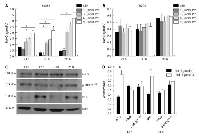
Figure 3 Prednisolone increases the release of nitric oxide in SaOS2.
A: SaOS2 were cultured in the presence of different concentrations of prednisolone. Nitric oxide was measured after 24, 48 and 96 h. Results are expressed as the mean ± SD of four different experiments (aP < 0.05, bP < 0.01, dP < 0.001); B: U2OS cells were processed as described in (A). No statistical significance was achieved; C: SaOS2 cells were exposed to prednisolone for 12 and 24 h and then lysed. 80 μg of protein extracts were loaded on SDS-PAGE. Western blots using specific antibodies against iNOS, eNOS, p-eNOS-P-Ser1177 were performed. Actin shows that equal amounts of protein were loaded per lane. The figure shows a representative blot; D: The histogram shows the quantitative evaluation of NOS/actin ratio by densitometry. Results are expressed as the mean ± SD of three separate experiments (bP < 0.01). CTR: Control; iNOS: Inducible nitric oxide synthase; eNOS: Endothelial nitric oxide synthase; Prd: Prednisolone.
- Citation: Cazzaniga A, Maier JAM, Castiglioni S. Prednisolone inhibits SaOS2 osteosarcoma cell proliferation by activating inducible nitric oxide synthase. World J Transl Med 2016; 5(1): 53-58
- URL: https://www.wjgnet.com/2220-6132/full/v5/i1/53.htm
- DOI: https://dx.doi.org/10.5528/wjtm.v5.i1.53


