
















Copyright: ©Author(s) 2026.
World J Transl Med. Nov 28, 2026; 12(4): 125141
Published online Nov 28, 2026. doi: 10.5528/wjtm.125141
Published online Nov 28, 2026. doi: 10.5528/wjtm.125141
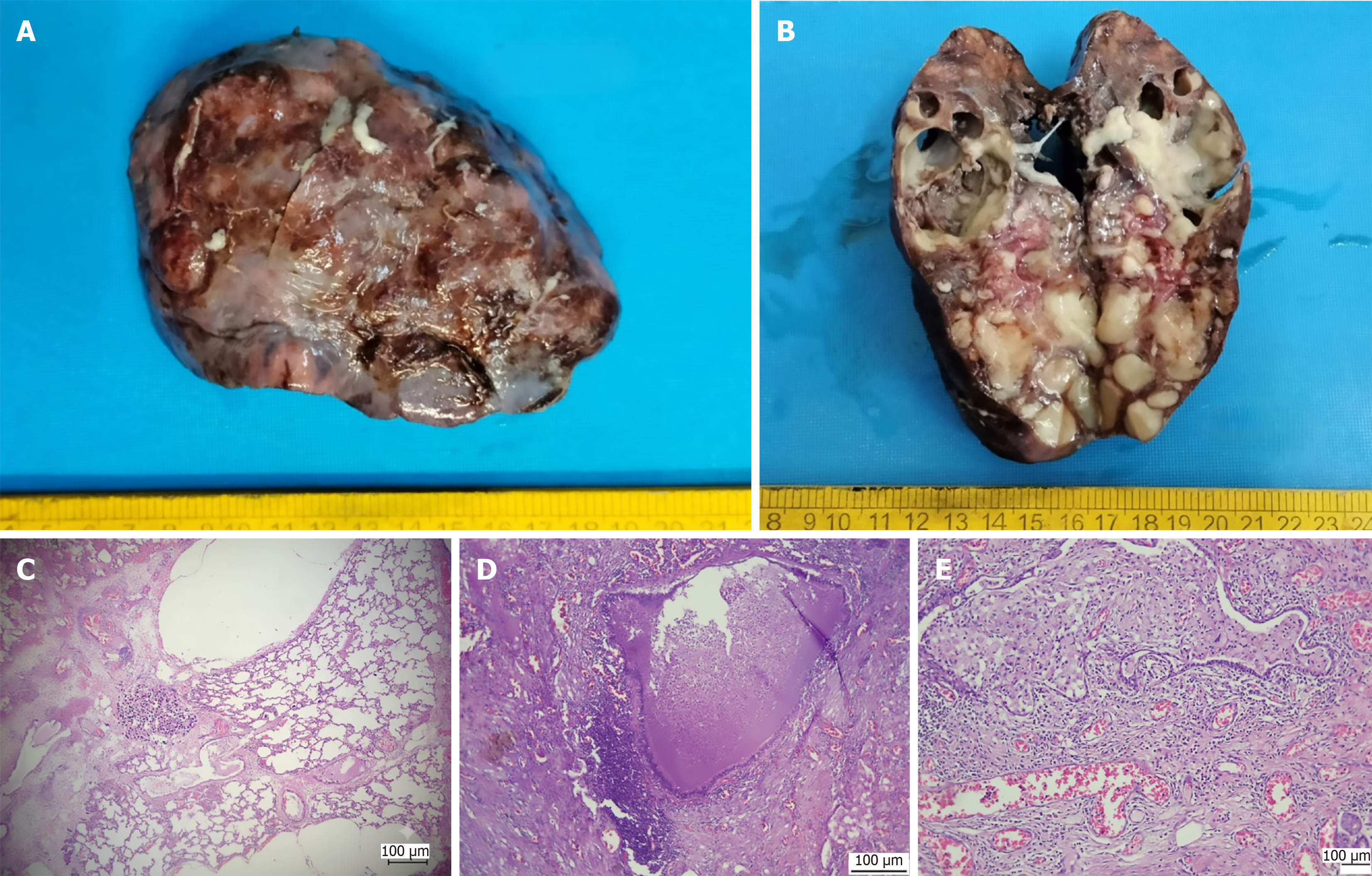
Figure 2 Specimen of right lower lobe of lung.
A: Externally the surface was irregular; B: Cut surface showed multiple cysts filled with pus and mucoid material; C and D: Histopathological features: Multiple blood vessels are seen with few appearing thick walled along with dilated cysts lined by ciliated columnar epithelium (4× magnification) (C) containing pools of mucin (mucostasis) (10× magnification), acute inflammation and necrosis (D); E: Dilated alveoli showed collection of foamy macrophages (10× magnification).
- Citation: Shah H, Patwardhan L, Sharma S. Congenital pulmonary airway malformation of a congenital lung disease with a potential hybrid lesion: A case report. World J Transl Med 2026; 12(4): 125141
- URL: https://www.wjgnet.com/2220-6132/full/v12/i4/125141.htm
- DOI: https://dx.doi.org/10.5528/wjtm.125141