Copyright
©2013 Baishideng Publishing Group Co.
World J Nephrol. Nov 6, 2013; 2(4): 111-124
Published online Nov 6, 2013. doi: 10.5527/wjn.v2.i4.111
Published online Nov 6, 2013. doi: 10.5527/wjn.v2.i4.111
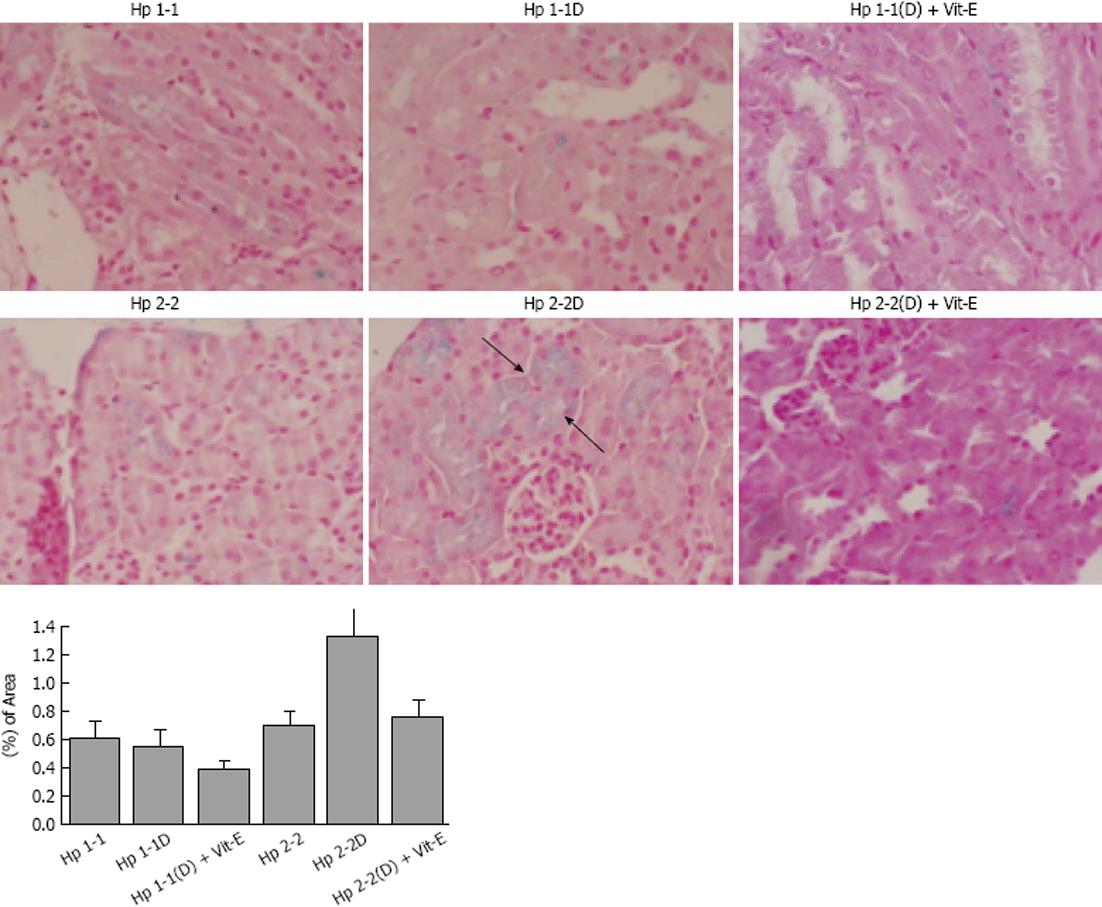
Figure 6 Increased renal iron deposition in the proximal tubule of haptoglobin 2-2 diabetes mellitus mice.
Perl’s iron stain was used to localize iron in paraffin-embedded kidney sections in haptoglobin (Hp) 1-1 and Hp 2-2 mice with and without diabetes mellitus (DM). Arrow indicates iron-induced stain in blue (× 400 magnification) located within proximal tubular cells. There was a significant increase in iron staining in the renal tissue of Hp 2-2 DM (D) vs Hp 1-1 DM (D) and Hp 2-2 non-DM mice (P < 0.001).
- Citation: Farid N, Inbal D, Nakhoul N, Evgeny F, Miller-Lotan R, Levy AP, Rabea A. Vitamin E and diabetic nephropathy in mice model and humans. World J Nephrol 2013; 2(4): 111-124
- URL: https://www.wjgnet.com/2220-6124/full/v2/i4/111.htm
- DOI: https://dx.doi.org/10.5527/wjn.v2.i4.111


