Copyright
©2013 Baishideng Publishing Group Co.
World J Nephrol. Nov 6, 2013; 2(4): 111-124
Published online Nov 6, 2013. doi: 10.5527/wjn.v2.i4.111
Published online Nov 6, 2013. doi: 10.5527/wjn.v2.i4.111
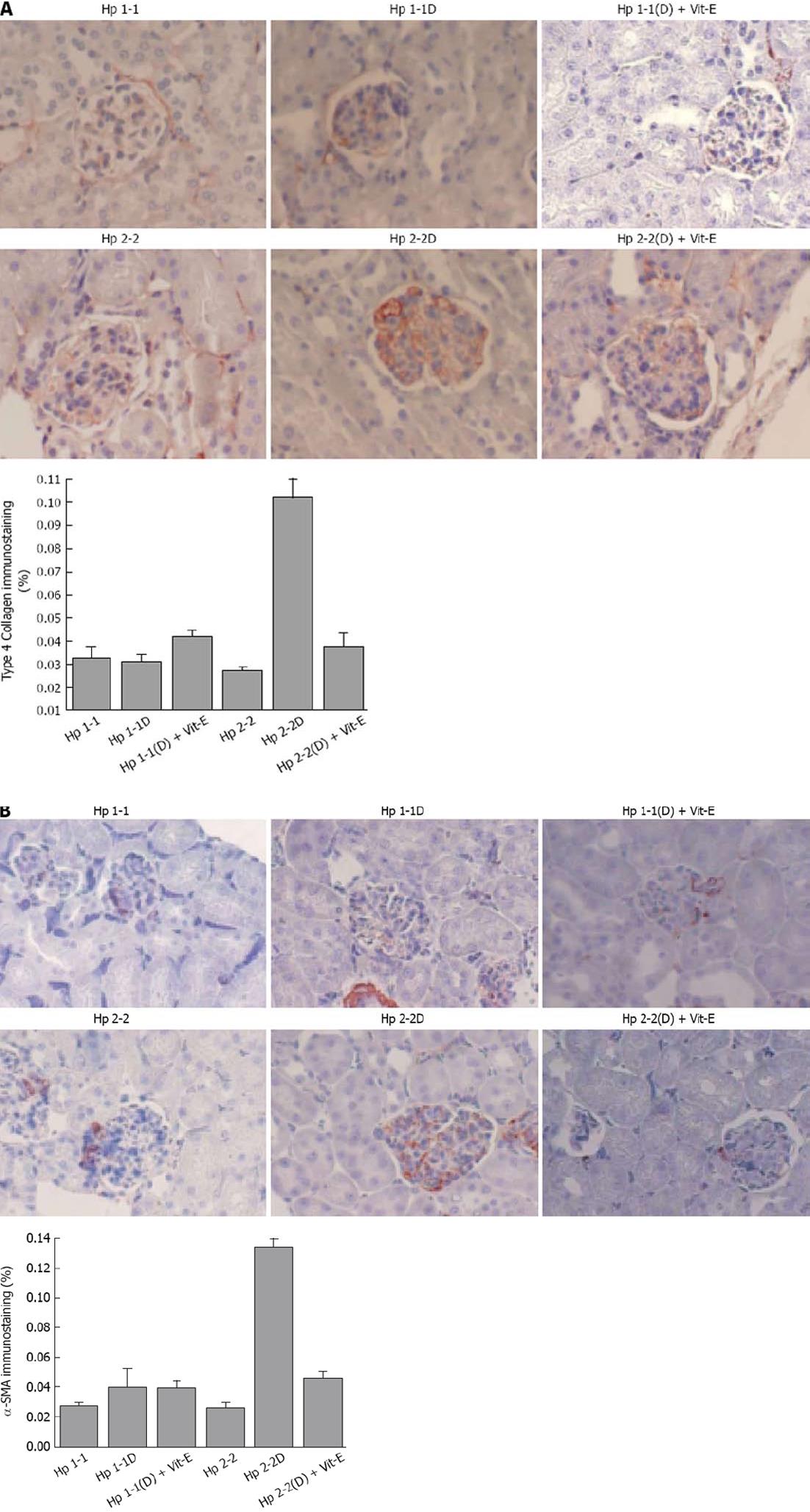
Figure 5 Increased mesangial collagen IV and smooth muscle actin in haptoglobin 2-2 diabetes mellitus mice.
A: Increased mesangial collagen IV in haptoglobin (Hp) 2-2 diabetes mellitus (DM) mice. Quantitation of the immunostaining area was reported in % area of the glomeruli. All values are expressed as means SE (30 glomeruli for each animal). There was a significant increase in collagen IV immunostaining (%) in Hp 2-2 DM vs Hp 1-1 DM mice (P < 0.001) and Hp 2-2 non-DM mice (P < 0.001). There was a significant decrease in collagen IV immunostaining area in Hp 2-2 DM mice with vitamin (Vit)-E (P < 0.05); B: Increased smooth muscle actin in Hp 2-2 DM mice. Immunohistochemical identification of smooth muscle actin (orange-red) was performed using a monoclonal antibody to mouse smooth muscle actin . All values are expressed as mean ± SE. Quantitation of actin staining was performed similar to collagen (as% of glomerular area) with a highly significant increase in actin staining in Hp 2-2 DM mice vs Hp 1-1 DM mice (P < 0.001) and Hp 2-2 non-DM mice (P < 0.001). There was a significant decrease in actin staining in Hp 2-2 DM mice treated with vitamin E (Vit-E) (P < 0.001).
- Citation: Farid N, Inbal D, Nakhoul N, Evgeny F, Miller-Lotan R, Levy AP, Rabea A. Vitamin E and diabetic nephropathy in mice model and humans. World J Nephrol 2013; 2(4): 111-124
- URL: https://www.wjgnet.com/2220-6124/full/v2/i4/111.htm
- DOI: https://dx.doi.org/10.5527/wjn.v2.i4.111


