
















Copyright: ©Author(s) 2026.
World J Nephrol. Mar 25, 2026; 15(1): 115252
Published online Mar 25, 2026. doi: 10.5527/wjn.v15.i1.115252
Published online Mar 25, 2026. doi: 10.5527/wjn.v15.i1.115252
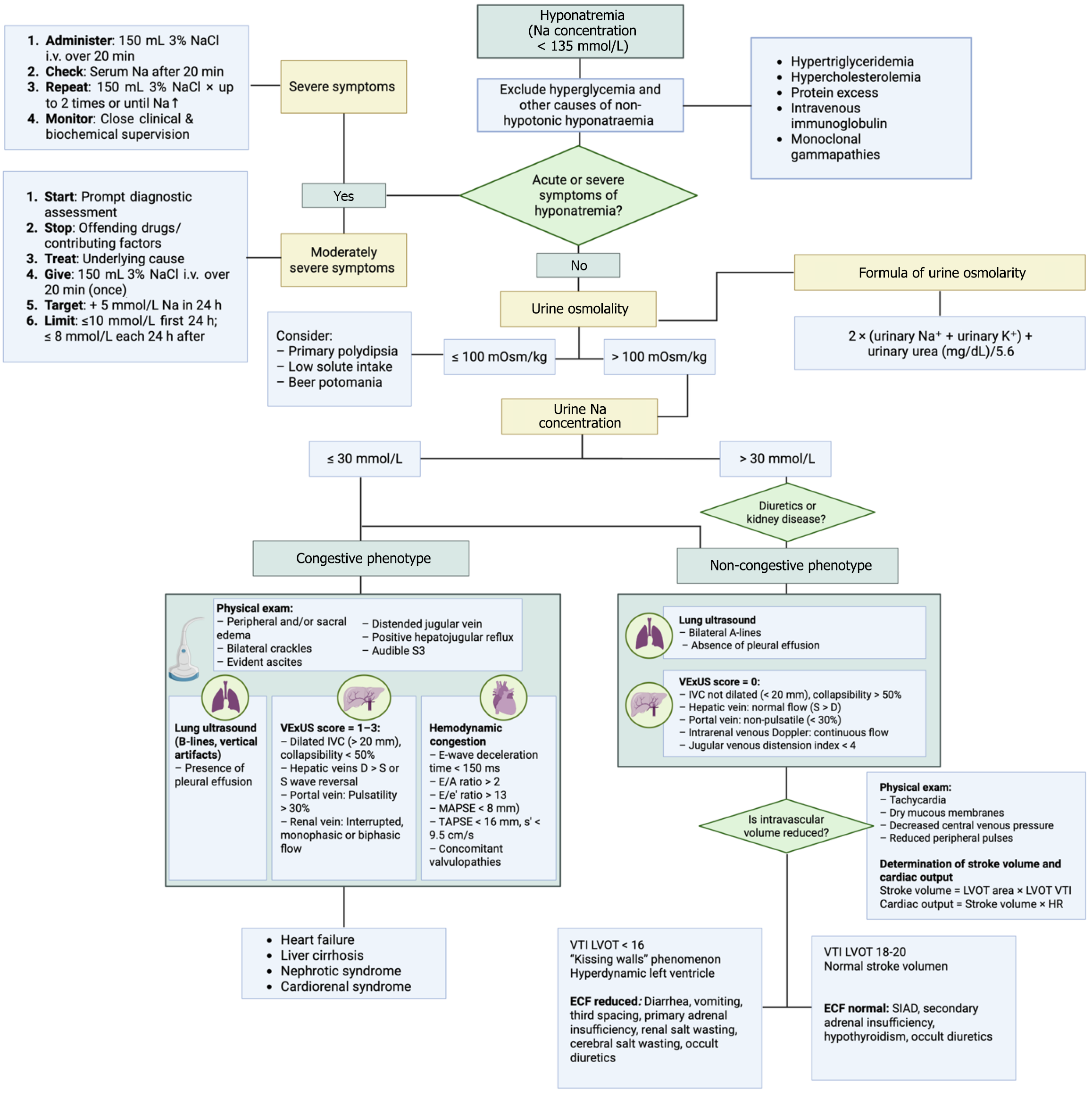
Figure 2 Diagnostic approach and phenotypic classification of hyponatremia.
Adapted from Eljazzar et al[38]. Algorithm outlining the stepwise evaluation of hyponatremia based on clinical severity, urine osmolality, and urine sodium concentration. A urinary sodium ≤ 30 mmol/L typically suggests low effective arterial volume; however, this does not always equate to true venous congestion. Differentiating congestive from non-congestive phenotypes requires a comprehensive assessment of volemic status, where point-of-care ultrasound plays a critical role in identifying hemodynamic congestion through evaluation of the inferior vena cava, hepatic, portal, and intrarenal venous flow patterns. IV: Intravenous; VExUS: Venous excess ultrasound; IVC: Inferior vena cava; E/A: Early to late diastolic transmitral flow velocity ratio; E/e’: Eatio of early mitral inflow velocity to early diastolic mitral annular velocity; MAPSE: Mitral annular plane systolic excursion; TAPSE: Tricuspid annular plane systolic excursion; LVOT: Left ventricular outflow tract; VTI: Velocity time integral; HR: Heart rate; ECF: Extracellular fluid; SIAD: Syndrome of inappropriate antidiuresis.
- Citation: Martínez-Sánchez FD, Gutierrez-Rosas LE, Barranco-Hernandez LH, Gonzalez-Alvarez G, Bastida-Castro LA, Rocha-Haro A, Barrientos-Cabrera G, Martínez-Cabrera CF, Balderas-Juarez J, Salinas-Ramirez MA, Hernandez-Castillo JL. Hyponatremia: Evolving diagnostics and emerging therapeutics in clinical practice. World J Nephrol 2026; 15(1): 115252
- URL: https://www.wjgnet.com/2220-6124/full/v15/i1/115252.htm
- DOI: https://dx.doi.org/10.5527/wjn.v15.i1.115252