Copyright
©The Author(s) 2025.
World J Nephrol. Jun 25, 2025; 14(2): 105374
Published online Jun 25, 2025. doi: 10.5527/wjn.v14.i2.105374
Published online Jun 25, 2025. doi: 10.5527/wjn.v14.i2.105374
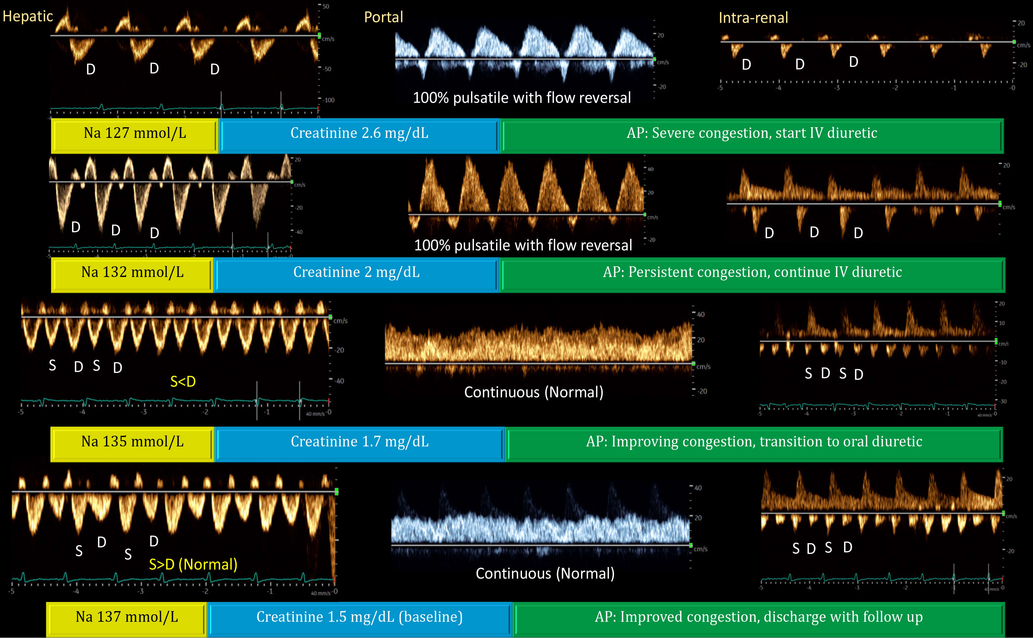
Figure 5 Doppler venous waveforms showing improvement (from top to bottom) in a patient with acute kidney injury and hyponatremia.
In a physiological state, the hepatic vein Doppler resembles the central venous waveform, with a larger S-wave compared to the D-wave. As right atrial pressure (RAP) increases, the S-wave amplitude decreases and eventually reverses, leaving only the D-wave below the baseline. Normally, the portal vein flow is continuous (less than 30% pulsatility), but pulsatility increases with rising RAP, eventually leading to late-systolic flow reversal (below-baseline flow). Increased portal vein pulsatility may indicate gut congestion, potentially affecting diuretic absorption. The renal parenchymal vein, typically continuous (like the portal vein but below the baseline), becomes more pulsatile with increasing RAP, eventually showing distinct S- and D-waves with S-reversal similar to the hepatic vein. Generally, improvements in the portal vein precede those in the hepatic and renal veins, as shown above. Renal interstitial edema may delay recovery of the venous waveform. Reproduced from Koratala et al[61]. AP: Assessment and plan; S-wave: Systolic wave; D-wave: Diastolic wave. Citation: Koratala A, Ronco C, Kazory A. Multi-Organ Point-Of-Care Ultrasound in Acute Kidney Injury. Blood Purif 2022; 51: 967-971. Copyright © 2022 S. Karger AG. Published by Karger Publishers. The authors have obtained the permission for figure using (Supplementary material).
- Citation: Diniz H, Ferreira F, Koratala A. Point-of-care ultrasonography in nephrology: Growing applications, misconceptions and future outlook. World J Nephrol 2025; 14(2): 105374
- URL: https://www.wjgnet.com/2220-6124/full/v14/i2/105374.htm
- DOI: https://dx.doi.org/10.5527/wjn.v14.i2.105374


