Copyright
©The Author(s) 2025.
World J Nephrol. Jun 25, 2025; 14(2): 103027
Published online Jun 25, 2025. doi: 10.5527/wjn.v14.i2.103027
Published online Jun 25, 2025. doi: 10.5527/wjn.v14.i2.103027
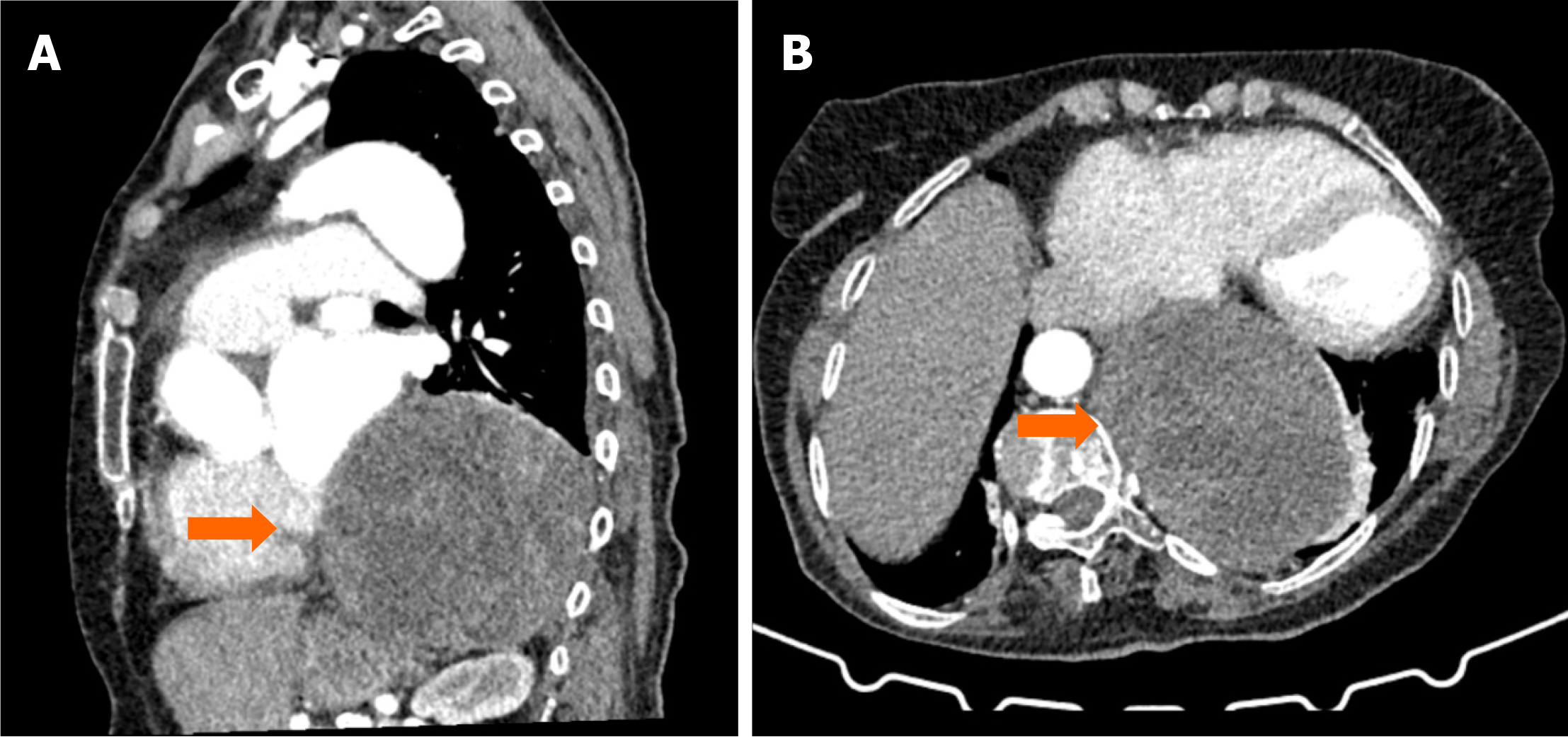
Figure 5 Thorax computed tomography images showing a cystic lesion in the left paravertebral area.
A: Sagittal contrast-enhanced thorax computed tomography (CT) scan demonstrates a cystic lesion with multiple enhancing septa and smooth contours, located extrapulmonary in the left paravertebral area (orange arrows); B: Axial contrast-enhanced thorax CT image further illustrates the extrapulmonary cystic lesion with well-defined septations (orange arrows).
- Citation: Celik AS, Yosunkaya H, Yayilkan Ozyilmaz A, Memis KB, Aydin S. Echinococcus granulosus in atypical localizations: Five case reports. World J Nephrol 2025; 14(2): 103027
- URL: https://www.wjgnet.com/2220-6124/full/v14/i2/103027.htm
- DOI: https://dx.doi.org/10.5527/wjn.v14.i2.103027


