Copyright
©The Author(s) 2025.
World J Nephrol. Jun 25, 2025; 14(2): 103027
Published online Jun 25, 2025. doi: 10.5527/wjn.v14.i2.103027
Published online Jun 25, 2025. doi: 10.5527/wjn.v14.i2.103027
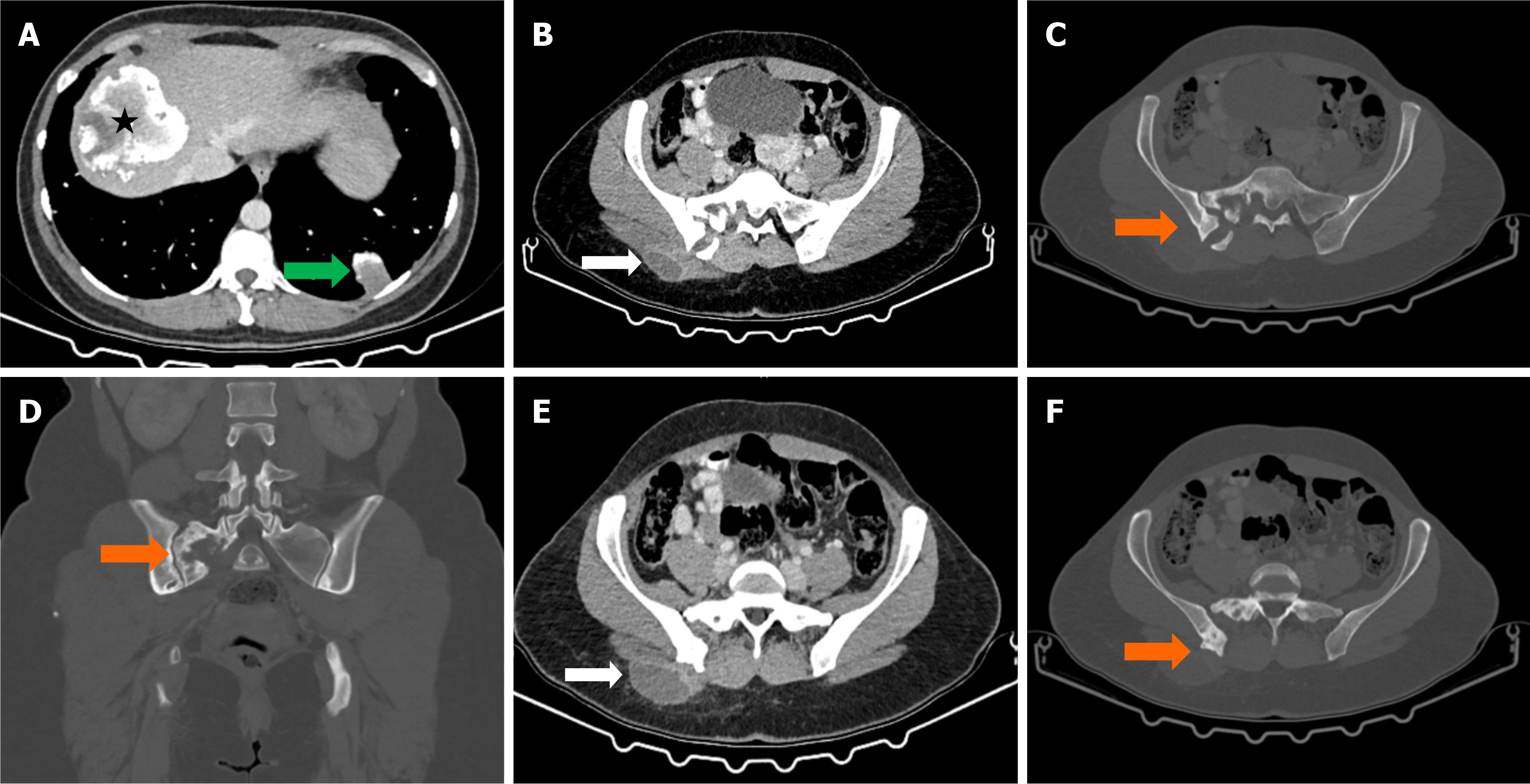
Figure 4 Abdominal computed tomography images showing inactive hydatid cysts and associated bone lesions.
A: Axial abdominal computed tomography (CT) image demonstrates an inactive hydatid cyst in the right lobe of the liver (black asterisk) and another inactive hydatid cyst in the lower lobe of the left lung (green arrow); B: Axial post-contrast abdominal CT image reveals a rim-enhancing cystic lesion in the subcutaneous adipose tissue of the right gluteal region (white arrows); C: Axial bone window CT image displays erosive lesions in the right half of the sacrum (orange arrows); D: Coronal bone window CT image highlights erosive lesions in the posterior part of the right iliac wing (orange arrows); E: Axial post-contrast abdominal CT image confirms rim enhancement in the gluteal cystic lesion; F: Axial bone window CT image further depicts bone erosions in the sacrum and iliac wing (orange arrows).
- Citation: Celik AS, Yosunkaya H, Yayilkan Ozyilmaz A, Memis KB, Aydin S. Echinococcus granulosus in atypical localizations: Five case reports. World J Nephrol 2025; 14(2): 103027
- URL: https://www.wjgnet.com/2220-6124/full/v14/i2/103027.htm
- DOI: https://dx.doi.org/10.5527/wjn.v14.i2.103027


