
















©The Author(s) 2024.
World J Nephrol. Sep 25, 2024; 13(3): 98300
Published online Sep 25, 2024. doi: 10.5527/wjn.v13.i3.98300
Published online Sep 25, 2024. doi: 10.5527/wjn.v13.i3.98300
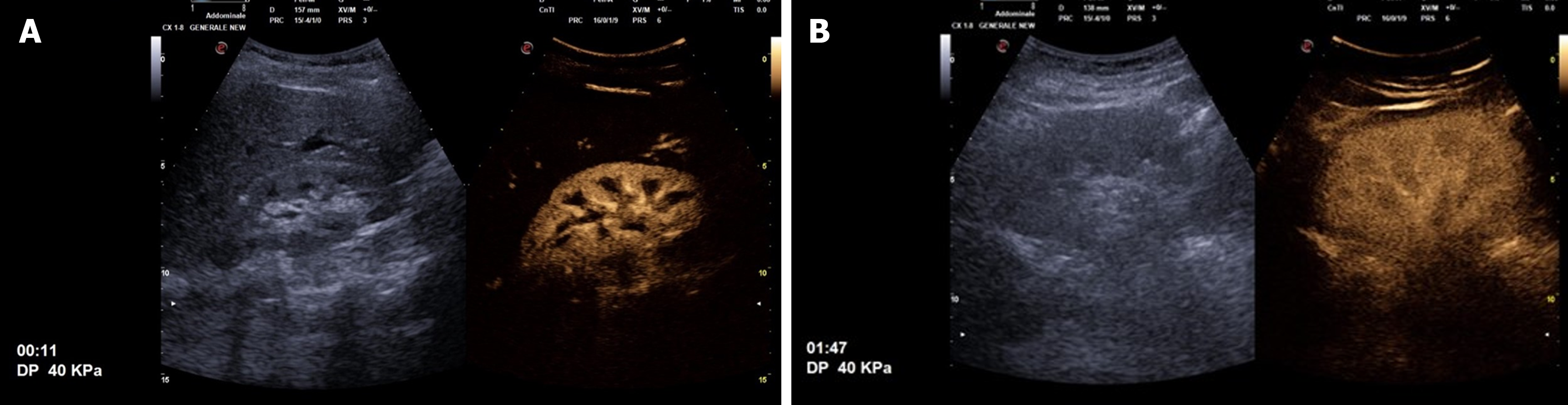
Figure 1 Following the ultrasound contrast agent injection.
A: Enhancement in the central arteries becomes visible 10-15 seconds after contrast injection, followed by the renal cortex a few seconds later, while the pyramids remain echo-poor; B: The renal pyramids gradually fill in, becoming almost isoechoic with the cortex within 30 seconds to 40 seconds after injection. During early-phase scanning, the kidneys appear hyperechoic compared to the liver or spleen. Later, the kidneys turn rapidly hypoechoic relative to the adjacent parenchyma, particularly the spleen.
- Citation: Boccatonda A, Stupia R, Serra C. Ultrasound, contrast-enhanced ultrasound and pyelonephritis: A narrative review. World J Nephrol 2024; 13(3): 98300
- URL: https://www.wjgnet.com/2220-6124/full/v13/i3/98300.htm
- DOI: https://dx.doi.org/10.5527/wjn.v13.i3.98300