Copyright
©The Author(s) 2019.
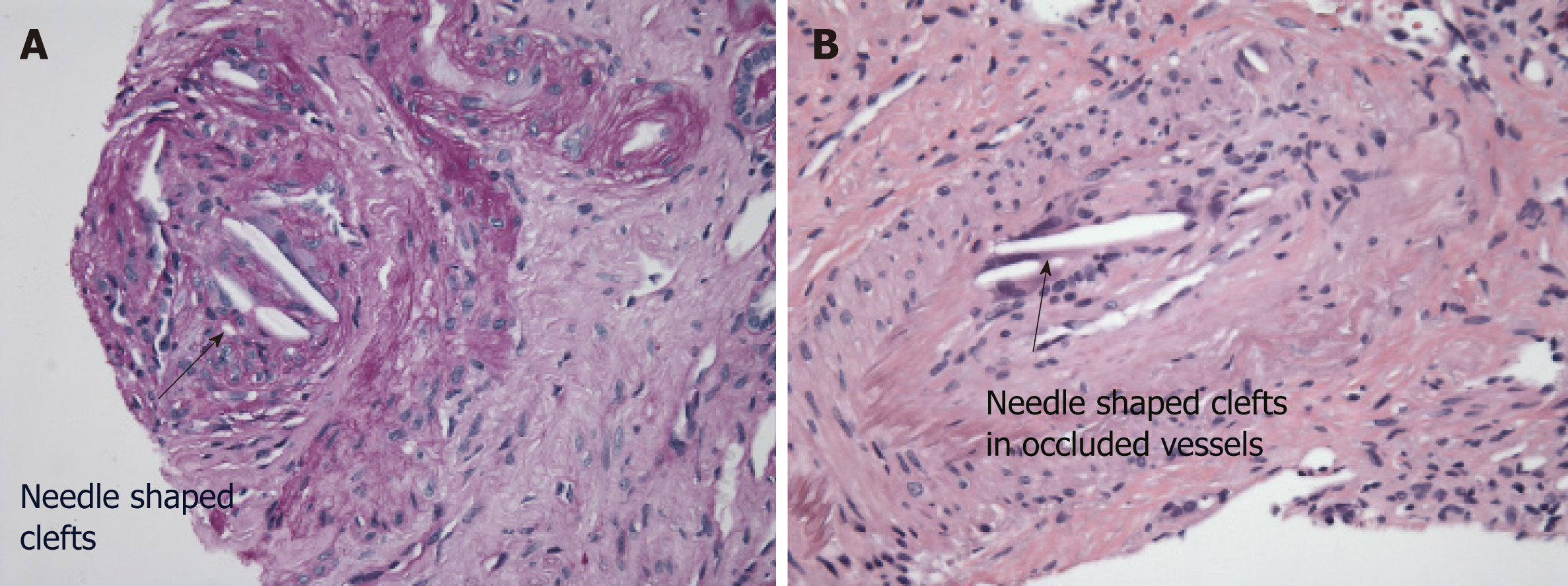
Figure 1 Atheroembolic disease of the kidney.
A, B: Several small arteries and arterioles are affected and show needle-shaped clefts in occluded and recanalized vessels (see arrow).
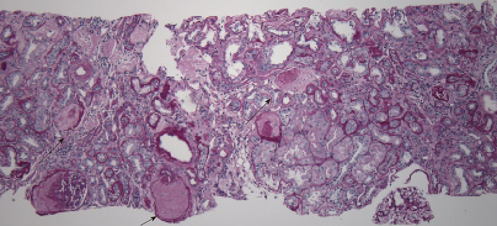
Figure 2 Focal global and segmental glomerulosclerosis (50% of glomeruli).
Arrow: Acute tubular injury, mild.
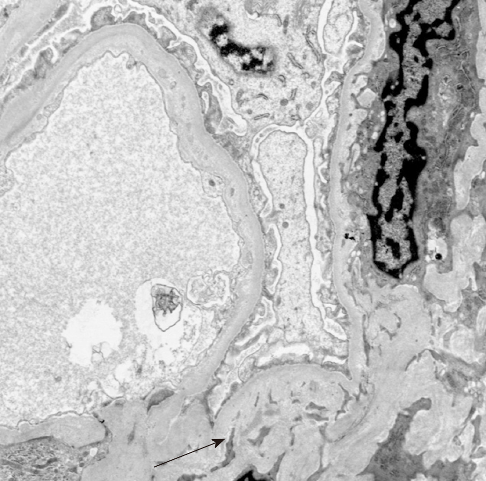
Figure 3 Electron microscopy showing sparsely distributed mesangial and glomerular capillary wall deposits (electron dense) (see arrow).
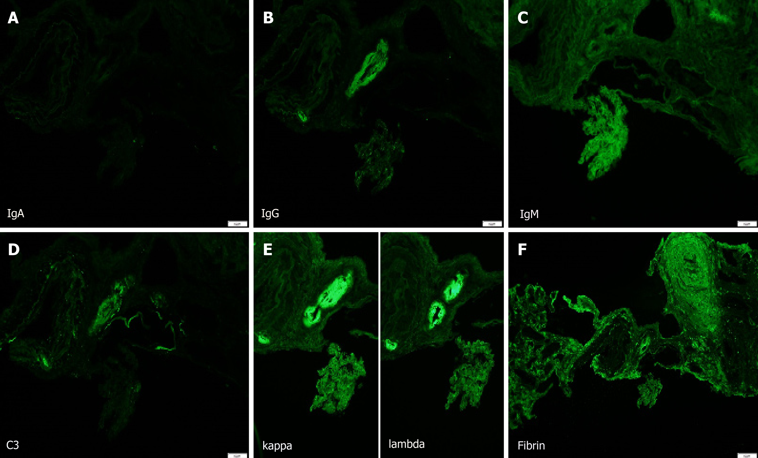
Figure 4 Ultra structural features suggestive of a mild and currently quiescent or inactive IgA nephropathy or sequelae of an IgA-dominant infection-associated glomerulonephritis.
There are sparsely distributed mesangial and glomerular capillary wall deposits reactive for IgA, IgM, C3, and with equal expression of kappa and lambda light chains. There are no signs of a currently active glomerulitis. A: Mild IgA deposition; B: IgG deposition; C: IgM deposition; D: C3 deposition; E: Kappa and lambda chain deposition; F: Fibrin deposition.
- Citation: Piranavan P, Rajan A, Jindal V, Verma A. A rare presentation of spontaneous atheroembolic renal disease: A case report. World J Nephrol 2019; 8(3): 67-74
- URL: https://www.wjgnet.com/2220-6124/full/v8/i3/67.htm
- DOI: https://dx.doi.org/10.5527/wjn.v8.i3.67


