
















©The Author(s) 2019.
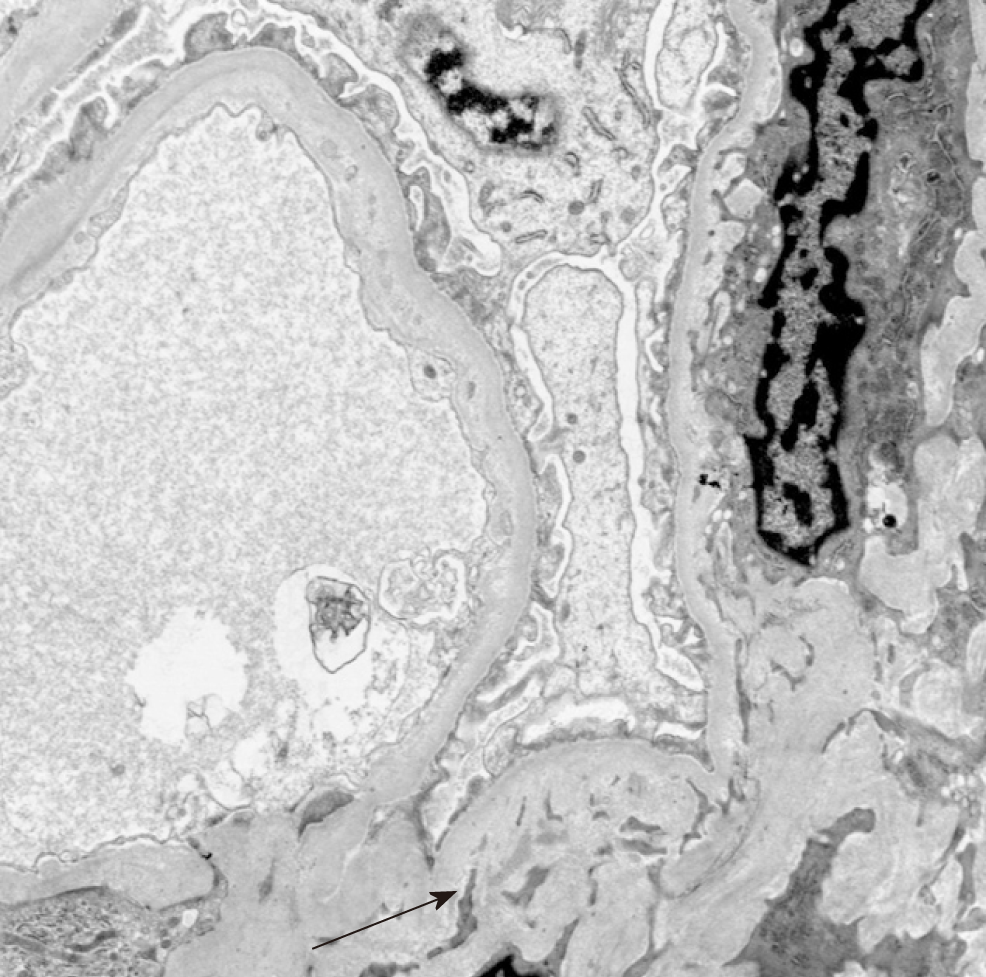
Figure 3 Electron microscopy showing sparsely distributed mesangial and glomerular capillary wall deposits (electron dense) (see arrow).
- Citation: Piranavan P, Rajan A, Jindal V, Verma A. A rare presentation of spontaneous atheroembolic renal disease: A case report. World J Nephrol 2019; 8(3): 67-74
- URL: https://www.wjgnet.com/2220-6124/full/v8/i3/67.htm
- DOI: https://dx.doi.org/10.5527/wjn.v8.i3.67