Copyright
©2013 Baishideng Publishing Group Co.
World J Virol. Aug 12, 2013; 2(3): 110-122
Published online Aug 12, 2013. doi: 10.5501/wjv.v2.i3.110
Published online Aug 12, 2013. doi: 10.5501/wjv.v2.i3.110
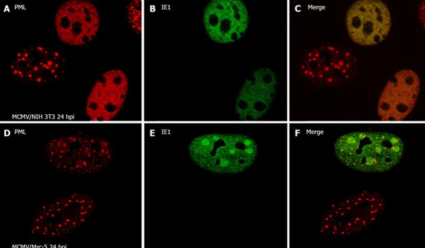
Figure 1 Immunofluorescent assay to show cytomegalovirus infection and nuclear domain 10.
A: After murine cytomegalovirus (MCMV) infection in NIH3T3 cells for 24 h, cells were stained with anti-Promyelocytic Leukemia bodies (PML) antibody (rabbit) to show nuclear domain 10 (ND10) (in red); B: Anti-IE1 antibody (mouse) was used to show IE1 (in green); C: The merged picture is shown in; D: After MCMV infection in Mrc-5 cells for 24 h, cells were stained with anti-PML antibody (rabbit) to show ND10 (in red); E: Anti-IE1 antibody (mouse) was used to show IE1 (in green); F: The merged picture is shown.
- Citation: Rivera-Molina YA, Martínez FP, Tang Q. Nuclear domain 10 of the viral aspect. World J Virol 2013; 2(3): 110-122
- URL: https://www.wjgnet.com/2220-3249/full/v2/i3/110.htm
- DOI: https://dx.doi.org/10.5501/wjv.v2.i3.110


