Copyright
©The Author(s) 2018.
World J Transplantation. Oct 22, 2018; 8(6): 203-219
Published online Oct 22, 2018. doi: 10.5500/wjt.v8.i6.203
Published online Oct 22, 2018. doi: 10.5500/wjt.v8.i6.203
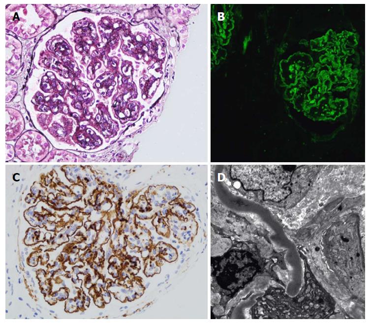
Figure 2 Renal histology in individuals with dense deposit disease.
A: Light microscopy with silver stain showing a membranoproliferative glomerulonephritis pattern with double contours of the glomerular basement membrane; B: Immunofluorescence; C: Immunohistochemistry with immunoperoxidase showing strong capillary wall staining of C3 and some granular mesangial C3; D: Characteristic sausage-like, intramembranous, osmiophilic deposits on electron microscopy. Adapted from Barbour et al[11].
- Citation: Abbas F, El Kossi M, Kim JJ, Shaheen IS, Sharma A, Halawa A. Complement-mediated renal diseases after kidney transplantation - current diagnostic and therapeutic options in de novo and recurrent diseases. World J Transplantation 2018; 8(6): 203-219
- URL: https://www.wjgnet.com/2220-3230/full/v8/i6/203.htm
- DOI: https://dx.doi.org/10.5500/wjt.v8.i6.203


