Copyright
©The Author(s) 2017.
World J Transplant. Dec 24, 2017; 7(6): 301-316
Published online Dec 24, 2017. doi: 10.5500/wjt.v7.i6.301
Published online Dec 24, 2017. doi: 10.5500/wjt.v7.i6.301
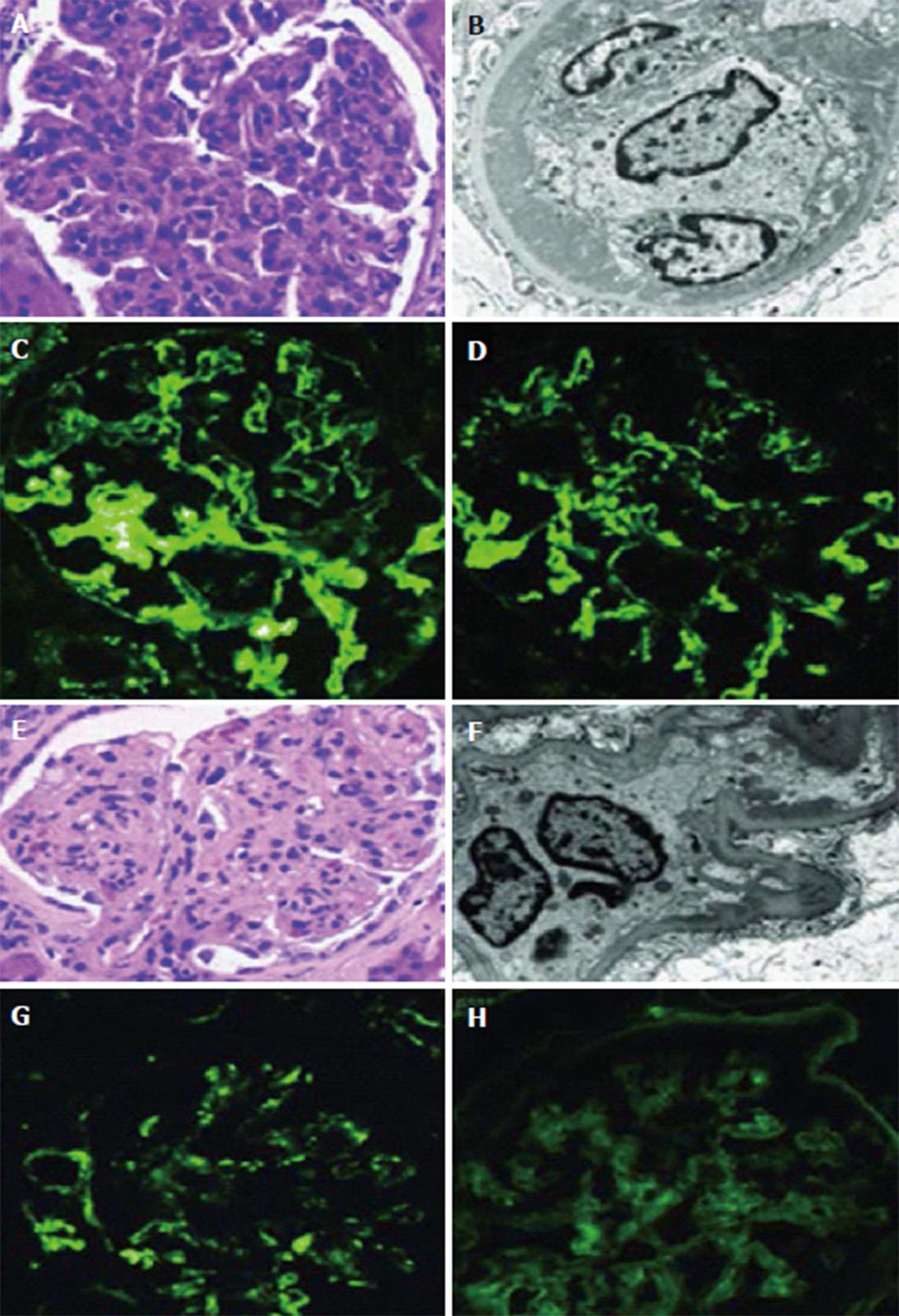
Figure 5 Histological changes of membranoproliferative glomerulonephritis in kidney transplant biopsies.
Typical LM, EM and IF finding in cases previously classified as MPGN. First panel shows a case reclassified as ICGN with C3 abnormalities, including (A) the classic MPGN pattern GN on LM (B) large sub-endothelial electron dense deposits on EM and granular mesangial and capillary wall staining for both (C) IgG and (D) C3 on IF. Second panel shows a case reclassified as a C3 glomerulopathy, with (E) a similar MPGN pattern on LM, (F) smaller sub-endothelial deposits on EM and granular mesangial and capillary wall staining for (G) C3, but no significant staining for (H) IgG. Adapted from Alasfar et al[30] with permission. LM: Light microscopic; EM: Electron microscopy; IF: Immunofluorescence.
- Citation: Abbas F, El Kossi M, Jin JK, Sharma A, Halawa A. Recurrence of primary glomerulonephritis: Review of the current evidence. World J Transplant 2017; 7(6): 301-316
- URL: https://www.wjgnet.com/2220-3230/full/v7/i6/301.htm
- DOI: https://dx.doi.org/10.5500/wjt.v7.i6.301


