Copyright
©The Author(s) 2025.
World J Transplant. Sep 18, 2025; 15(3): 104109
Published online Sep 18, 2025. doi: 10.5500/wjt.v15.i3.104109
Published online Sep 18, 2025. doi: 10.5500/wjt.v15.i3.104109
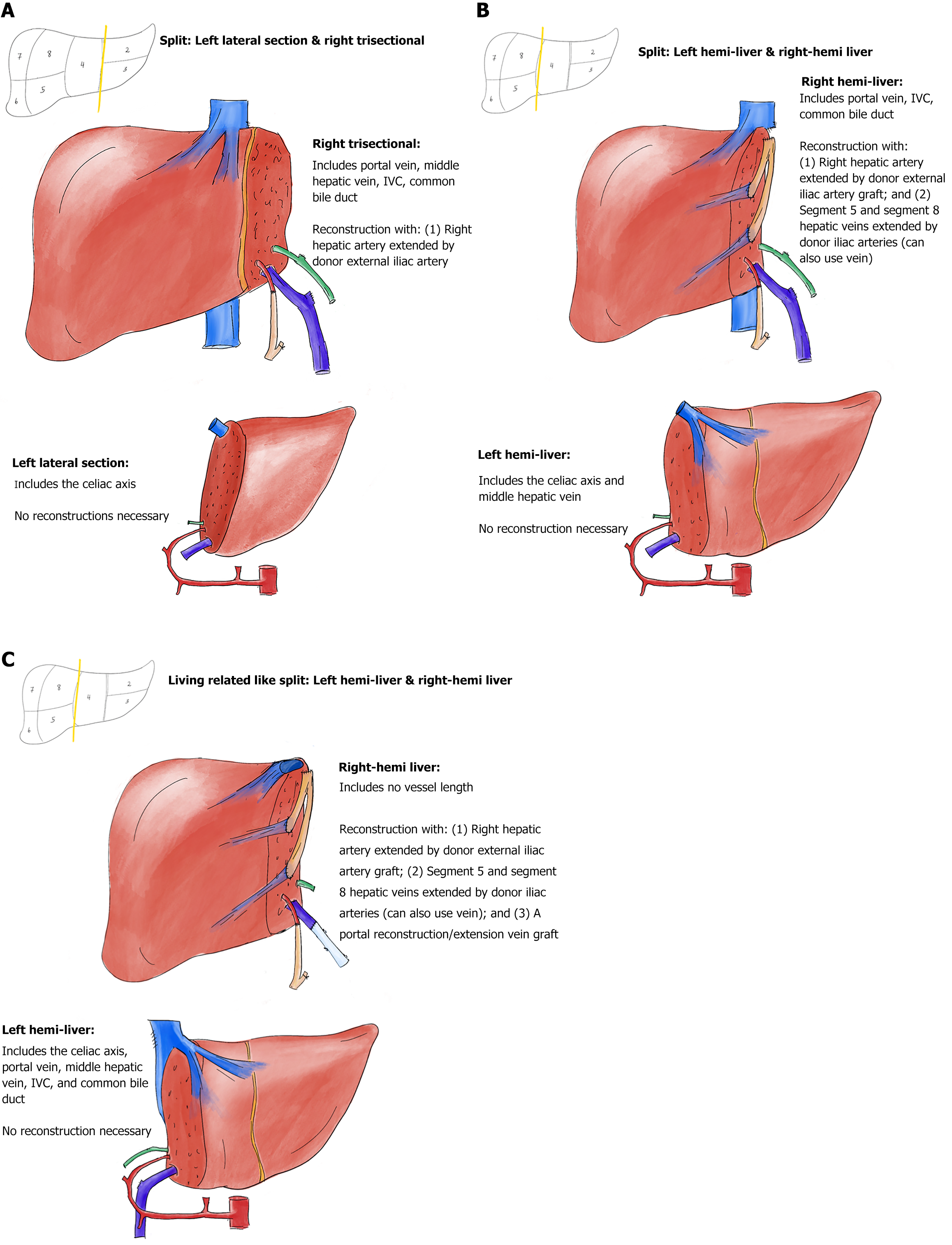
Figure 1 Deceased donor partial graft variants with vascular reconstructions.
A: Depiction of a left lateral–right trisectional graft split; B: Depiction of a left-right hemi-liver graft split, with the inferior vena cava (IVC) staying with the right hemi-liver graft; C: Depiction of a left-right hemi-liver graft split with the IVC staying with the left hemi-liver graft (living related donor like). All vascular inflow and outflow reconstructions in the depictions are performed before the connection to the normothermic machine perfusion pump, except the segments 5 and 8 hepatic veins outflow restorations in Figure 1C. Arteries are depicted in red; arterial grafts are depicted in orange; bile ducts are depicted in green; veins are depicted in dark blue; vein grafts are depicted in light blue.
- Citation: Baimas-George M, Archie WH, Soltys K, Soto JR, Levi D, Eskind L, Casingal V, Denny R, Attia M, Mazariegos GV, Vrochides D. Optimizing liver utilization for transplantation with partial grafts undergoing normothermic machine perfusion: Two case reports. World J Transplant 2025; 15(3): 104109
- URL: https://www.wjgnet.com/2220-3230/full/v15/i3/104109.htm
- DOI: https://dx.doi.org/10.5500/wjt.v15.i3.104109


