









BPG is committed to discovery and dissemination of knowledge
Review
Copyright: ©Author(s) 2026.
World J Psychiatry. Nov 19, 2026; 16(11): 125256
Published online Nov 19, 2026. doi: 10.5498/wjp.125256
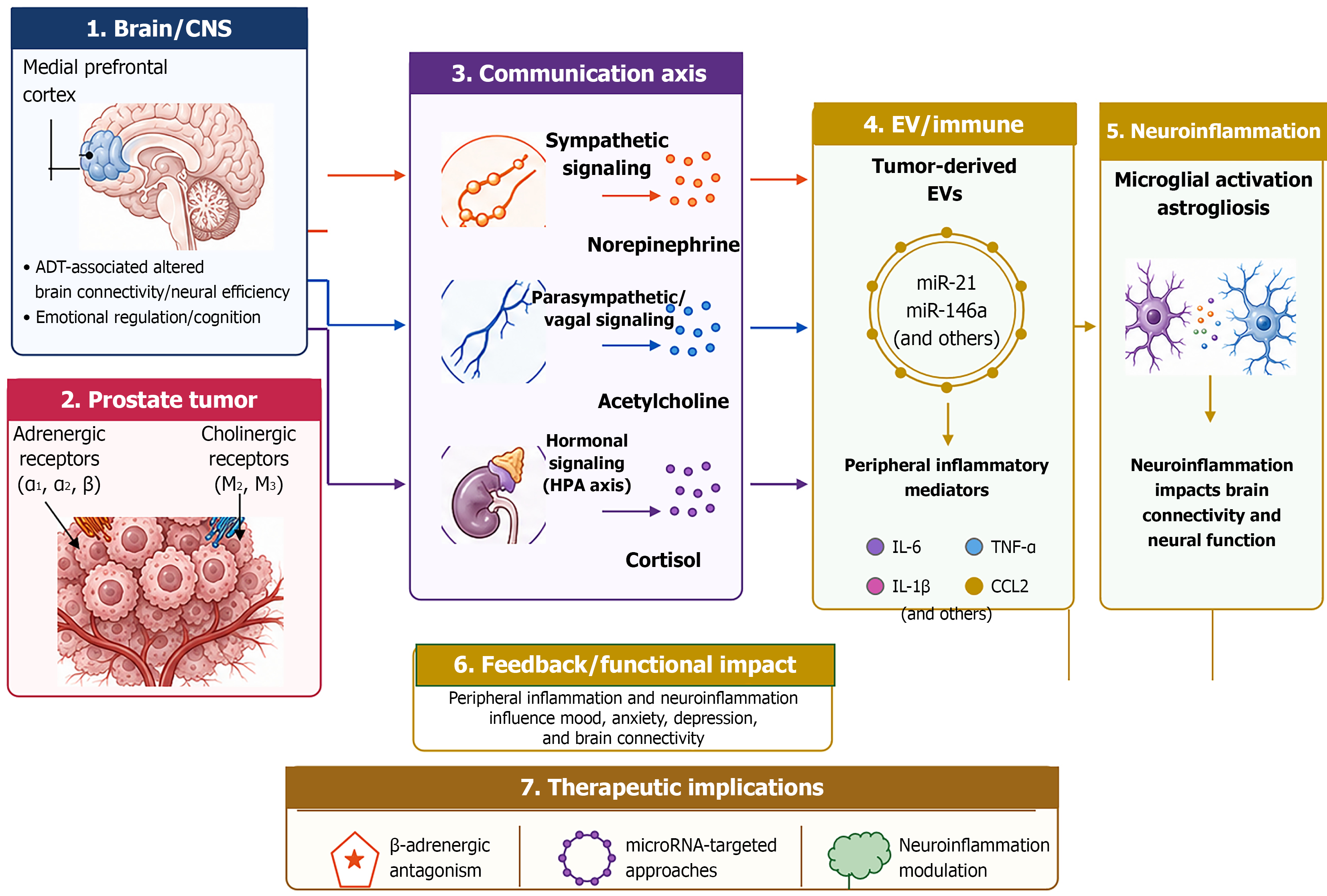
Figure 2
Figure 2 Depicts the brain-tumor communication through autonomic, hormonal, extracellular-vesicle, and inflammatory signaling in prostate cancer. Author-assembled schematic illustrating likely bidirectional communication pathways between the prostate tumor, systemic signalling and the brain. Published human data in prostate cancer (PCa) include observations of decreased medial prefrontal activation with a relationship between Medial P-F activation and connectivity and treatment with androgen deprivation therapy[70,92], as well as altered systemic inflammation and symptom burden during treatment[98], a pilot of neuroinflammation in androgen deprivation therapy recipients[42] and circulating PCa extracellular vesicle miRNAs, like miR-21-5p[99]. From PCa cell and animal studies come evidence that autonomic innervation regulates tumor function[78] and the role of exosomal miR-146a-5p signaling within the prostate tumor environment[100]. Asterisked neuroinflammatory links – including microglial activation, astrogliosis, and their relationship with behavioral or neural effects – have not been established in PCa patients and are extrapolated primarily from non-prostate-cancer tumor-bearing animal models[101,102]. The therapeutic implications shown are exploratory research directions rather than established psycho-oncology interventions; β-blocker evidence in PCa is observational rather than interventional[96]. CNS: Central nervous system; ADT: Androgen deprivation therapy; HPA: Hypothalamic-pituitary-adrenal; EVs: Extracellular vesicles; IL: Interleukin; TNF-α: Tumor necrosis factor alpha; CCL2: Chemokine (C-C motif) ligand 2.


Write to the Help Desk